. 2022 Dec 16:10:1049678.
doi: 10.3389/fcell.2022.1049678. eCollection 2022.

Sustained therapeutic effect of an anti-inflammatory peptide encapsulated in nanoparticles on ocular vascular leakage in diabetic retinopathy

Affiliations

Sustained therapeutic effect of an anti-inflammatory peptide encapsulated in nanoparticles on ocular vascular leakage in diabetic retinopathy

Qiang Qu et al. Front Cell Dev Biol. .

Abstract

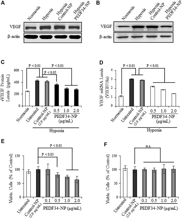
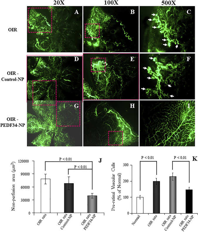
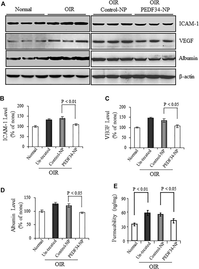
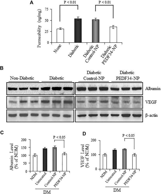
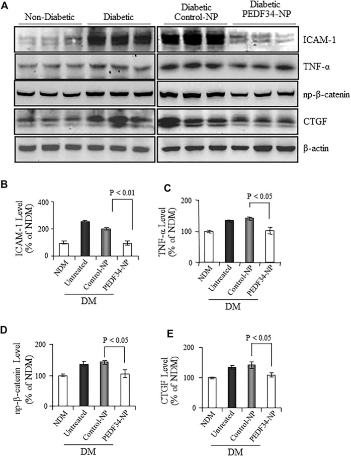
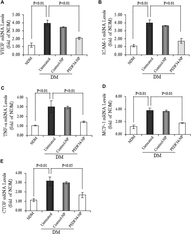
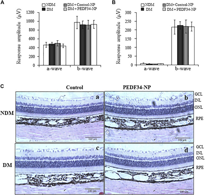
Pigment epithelium-derived factor (PEDF), an endogenous Wnt signaling inhibitor in the serine proteinase inhibitors (SERPIN) super family, is present in multiple organs, including the vitreous. Significantly low levels of PEDF in the vitreous are found to associate with pathological retinal vascular leakage and inflammation in diabetic retinopathy (DR). Intravitreal delivery of PEDF represents a promising therapeutic approach for DR. However, PEDF has a short half-life after intravitreal injection, which represents a major hurdle for the long-term treatment. Here we report the prolonged therapeutic effects of a 34-mer peptide of the PEDF N-terminus, encapsulated in poly (lactic-co-glycolic acid) (PLGA) nanoparticles (PEDF34-NP), on DR. PEDF34-NP inhibited hypoxia-induced expression of vascular endothelial growth factor and reduced levels of intercellular adhesion molecule 1 (ICAM-1) in cultured retinal cells. In addition, PEDF34-NP significantly ameliorated ischemia-induced retinal neovascularization in the oxygen-induced retinopathy rat model, and significantly reduced retinal vascular leakage and inflammation in streptozotocin-induced diabetic rats up to 4 weeks after intravitreal injection, as compared to PLGA-NP control. Intravitreal injection of PEDF34-NP did not display any detectable toxicities to retinal structure and function. Our findings suggest that PEDF34-NP can confer sustained therapeutic effects on retinal inflammation and vascular leakage, having considerable potential to provide long-term treatment options for DR.

Keywords: PLGA; anti-inflammatory; anti-vascular leakage; diabetic retinopathy; nanoparticles; pigment epithelium-derived factor; wnt signaling.


Conflict of interest statement

DW, RF, was employed by EyeCro LLC. The remaining authors declare that the research was conducted in the absence of any commercial or financial relationships that could be construed as a potential conflict of interest.

Figures

FIGURE 1
FIGURE 1
Inhibitory effect of PEDF34-NP on VEGF expression induced by hypoxia in ARPE19 cells and BRCEC. (A) ARPE19 cells were grown to 70% confluence in media containing 2% FBS and treated with PEDF34-NP at 1 μg/ml for 16 h under hypoxia (1% oxygen). The same amount of total cellular proteins (30 µg) was applied for Western blot analysis using the antibody specific for VEGF. (B) BRCEC were exposed to hypoxia and PEDF-34NP for 16 h. VEGF levels in the cells were measured by Western blot analysis. (C) and (D) ARPE19 cells were treated with Control-NP and various concentrations of PEDF34-NP for 24 h under hypoxia. VEGF secreted into the media (sVEGF) was measured by ELISA using an ELISA kit (C) and VEGF mRNA levels were measured by real-time RT-PCR and normalized by 18S rRNA levels (D). (E) and (F) BRCEC (E) and ARPE19 cells (F) were treated with PEDF34-NP at the indicated concentrations for 24 h. Viable cells were quantified using the MTT assay. All values are mean ± SD (n = 3).
FIGURE 2
FIGURE 2
Inhibitory effects of PEDF34-NP on ischemia-induced retinal neovascularization. Rats were exposed to 75% ± 2% oxygen from P7 to P12. Retinal vasculature was examined using fluorescein angiography at P18 as described in Methods. (A–C) OIR rats without treatment; (D–F) OIR rats injected with Control-NP (5 µg/eye); (G–I) OIR rats injected with PEDF34-NP into the vitreous (5 µg/eye) at P12. (J) Non-perfusion area was measured and expressed as percent of the total retina area (mean ± SD, n = 6). (K) At P18, the eyes were fixed, sectioned and stained with hematoxylin and eosin (H&E). Pre-retinal vascular cells were counted in eight non-continuous sections per eye and averaged as described in Methods (mean ± SD, n = 6).
FIGURE 3
FIGURE 3
Effect of PEDF34-NP on retinal inflammatory factor expression and vascular leakage in OIR rats. OIR rats received an intravitreal injection of 5 µg PEDF34-NP or Control-NP. (A) At P16 the retina was perfused, and retinal levels of ICAM-1, VEGF, and albumin were determined by western blot analysis, with β-actin as loading control. (B–D) Retinal levels of ICAM-1, VEGF, and albumin were quantified by densitometry and expressed as % of that in normal rat retinas (mean ± SD, n = 3). (E) Retinal vascular permeability was measured using the Evans blue as tracer at P16, and normalized by the total retinal protein concentration (mean ± SD, n = 6).
FIGURE 4
FIGURE 4
Effect of PEDF34-NP on retinal vascular leakage in STZ-induced diabetic rats. Diabetic rats received an intravitreal injection of 5 μg PEDF34-NP or Control-NP. (A) Four weeks after the injection, retinal vascular permeability was measured using Evans blue as tracer and normalized by total retinal protein concentration (mean ± SD, n = 5). (B–D) Retinas were dissected after perfusion, and the levels of albumin and VEGF were determined by western blot analysis, normalized to β-actin levels, semi-quantified by densitometry, and expressed as % of those in non-diabetic rat retinas (mean ± SD, n = 3).
FIGURE 5
FIGURE 5
PPEDF34-NP decreased retinal levels of non-phosphorylated β-catenin (NP-β-catenin), inflammatory, and fibrogenic factors in STZ-induced diabetic rats. Diabetic rats received an intravitreal injection of 5 μg of PEDF34-NP or Control-NP. (A) Four weeks after the injection, retinal levels of ICAM-1, TNF-α NP-β-catenin, and CTGF were determined by western blot analysis. PEDF34-NP significantly reduced the levels of ICAM-1 (B), TNF-α (C), NP-β-catenin (D), and CTGF (E) in the retinal tissue. The proteins levels were semi-quantified by densitometry, normalized by β-actin levels, and expressed as % of those in non-diabetic rat retinas (mean ± SD, n=3).
FIGURE 6
FIGURE 6
PEDF34-NP treatment decreased mRNA levels of inflammatory and fibrogenic factors in the STZ-induced diabetic rats. Diabetic rats received an intravitreal injection of 5 μg of PEDF34-NP or Control-NP. RNA was extracted from the retina 4 weeks after the injection, and mRNA levels of Vegf (A) , Icam-1 (B) , Tnf-α (C), Mcp-1 (D), and Ctgf (E) were semi-quantified by real-time RT-PCR and expressed as fold of that in non-diabetic rat retinas (mean ± SD, n = 3).
FIGURE 7
FIGURE 7
No changes in ERG response and retinal histology in response to PEDF34-NP injection. Scotopic and photopic ERG were recorded from six rats at 4 weeks after the intravitreal injection of PEDF34-NP and Control-NP as indicated. Amplitudes of a- and b-waves from scotopic (A) and photopic (B) ERG were averaged and compared (mean ± SD, n = 6). (C) Representative images of retinal section stained with H&E from non-diabetic rats (A), non-diabetic rats (NDM) injected with PEDF34-NP (B), diabetic rats (DM) (C), and diabetic rats injected with PEDF34-NP (D).

Similar articles

Cited by

References

    1. Abramson L. P., Stellmach V., Doll J. A., Cornwell M., Arensman R. M., Crawford S. E. (2003). Wilms' tumor growth is suppressed by antiangiogenic pigment epithelium-derived factor in a xenograft model. J. Pediatr. Surg. 38, 336–342. discussion 336-42. 10.1053/jpsu.2003.50104 - DOI - PubMed
    1. Adamis A. P., Miller J. W., Bernal M. T., D'Amico D. J., Folkman J., Yeo T. K., et al. (1994). Increased vascular endothelial growth factor levels in the vitreous of eyes with proliferative diabetic retinopathy. Am. J. Ophthalmol. 118, 445–450. 10.1016/s0002-9394(14)75794-0 - DOI - PubMed
    1. Adamis A. P., Shima D. T., Tolentino M. J., Gragoudas E. S., Ferrara N., Folkman J., et al. (1996). Inhibition of vascular endothelial growth factor prevents retinal ischemia-associated iris neovascularization in a nonhuman primate. Arch. Ophthalmol. 114, 66–71. 10.1001/archopht.1996.01100130062010 - DOI - PubMed
    1. Adamis A. P., Aiello L. P., D'Amato R. A. (1999). Angiogenesis and ophthalmic disease. Angiogenesis 3, 9–14. 10.1023/a:1009071601454 - DOI - PubMed
    1. Ahmadi M. A., Lim J. I. (2009). Update on laser treatment of diabetic macular edema. Int. Ophthalmol. Clin. 49, 87–94. 10.1097/IIO.0b013e31819fd6b2 - DOI - PubMed