Review
. 2022 Dec 16:13:1079217.
doi: 10.3389/fendo.2022.1079217. eCollection 2022.

Classification of diabetic retinopathy: Past, present and future

Affiliations
Review

Classification of diabetic retinopathy: Past, present and future

Zhengwei Yang et al. Front Endocrinol (Lausanne). .

Abstract

Diabetic retinopathy (DR) is a leading cause of visual impairment and blindness worldwide. Since DR was first recognized as an important complication of diabetes, there have been many attempts to accurately classify the severity and stages of disease. These historical classification systems evolved as understanding of disease pathophysiology improved, methods of imaging and assessing DR changed, and effective treatments were developed. Current DR classification systems are effective, and have been the basis of major research trials and clinical management guidelines for decades. However, with further new developments such as recognition of diabetic retinal neurodegeneration, new imaging platforms such as optical coherence tomography and ultra wide-field retinal imaging, artificial intelligence and new treatments, our current classification systems have significant limitations that need to be addressed. In this paper, we provide a historical review of different classification systems for DR, and discuss the limitations of our current classification systems in the context of new developments. We also review the implications of new developments in the field, to see how they might feature in a future, updated classification.

Keywords: artificial intelligence; classification; deep learning; diabetic retinopathy; imaging technology; pathophysiology; quantitative assessment; severity staging system.


Conflict of interest statement

The authors declare that the research was conducted in the absence of any commercial or financial relationships that could be construed as a potential conflict of interest.

Figures

Figure 1
Figure 1
Pathophysiology of diabetic retinopathy Hyperglycaemia cascade of events leading to neurodegeneration and microvascular impairment, which are the two key main pathways to result in the development of diabetic retinopathy. Neurodegeneration can be activated by glutamate excitotoxicity, loss of neuroprotection, and impairment of neurovascular coupling. Meanwhile, impairment of neurovascular coupling can lead to microvascular impairment, which can trigger the formation of DME and retinal neovascularization. AGE, advanced glycation end-products; RAS, renin-angiotensin system; PKC, protein kinase C; DME, diabetic macular edema; CA, carbonic anhydrase; VEGF, vascular endothelial growth factor; EPO, erythropoietin; GH-IGF, growth hormone-insulin growth factor.
Figure 2
Figure 2
Optical coherence tomography (OCT) signs of diabetic macular edema (DME). (A) All retinal layers are intact and visible. The retinal profile is not altered. But there is diffuse macular thickening. (B) Vitreomacular traction with a thick posterior hyaloid membrane (white arrowheads), small cystoid spaces (oblique white arrowheads) and hard exudates (black asterisk) in the outer plexiform layer and the outer nuclear layer. (C) Multiple hyperreflective retinal foci (HRF) are seen. Subretinal fluid causing a neurosensory detachment of the fovea (white asterisk). (D) Cystic cavities, hard exudates, and HRF located in the outer retina, but the external limiting membrane (ELM) and ellipsoid zone (EZ) are intact. (E) The magnified image (white square) shows the bridging retinal processes (white arrowheads) between the cystic cavities. (F) Multiple cystoid spaces and HRF in the inner and outer layers with disorganization of the inner retinal layers (DRIL; white bracket in the magnified image). The ELM and EZ are disrupted under the fovea.
Figure 3
Figure 3
Comparison of an ultra-wide field (UWF) retinal image and the Early Treatment Diabetic Retinopathy Study (ETDRS) 7 standard photographic fields. UWF retinal image is superimposed by the ETDRS 7 standard fields in white circles. The white arrowheads showing diabetic retinopathy lesions predominantly peripheral to the ETDRS fields.
Figure 4
Figure 4
Comparison of paired standard 45° fundus photographs and ultra-widefield photographs in three diabetic patients. (A, B), Standard 45° fundus photograph and ultra-widefield photograph from the left eye of the same patient, with no diabetic retinopathy. (C, D), Standard 45° fundus photograph showing microaneurysms, hard exudate, cotton wool spots and dot-blot retinal hemorrhages from diabetic retinopathy in the posterior pole, and accompanying ultra-widefield photograph from the same eye showing more retinal lesions in the periphery. (E, F), Standard 45° fundus photograph showing an eye with diabetic retinopathy that has undergone panretinal laser photocoagulation, and the accompanying ultra-widefield photograph from the same eye showing the peripheral extent of the laser photocoagulation scars. (a) microaneurysms, (b) hemorrhage, (c) hard exudate, (d) cotton wool spots, and (e) photocoagulation scars.
Figure 5
Figure 5
Multimodal images of proliferative diabetic retinopathy in both eyes of the same patient. Ultra-wide field (UWF) retinal images with the ETDRS 7-feld, 30-degree fundus images in circles outlined in white. The UWF fundus imaging of right eye (A) and left eye (B) showing retinal hemorrhages, microaneurysms, hard exudates, cotton wool spots, abnormal vascular loop, intraretinal microvascular abnormalities (IRMA), and retinal neovascularization. The ultra-widefield fluorescein angiography of right eye (C) and left eye (D) illustrating the corresponding hyperfluorescent dots of microaneurysms, areas of capillary non-perfusion, and multiple small areas of neovascularization identified by the hyperfluorescent leakage of dye. Corresponding wide field swept-source optical coherence tomography angiography (WF SS-OCTA) of right eye (E) and left eye (F) exhibiting area of non-perfusion, abnormal vascular loop, IRMA, and retinal neovascularization. (a) microaneurysms, (b) hemorrhage, (c) hard exudates, (d) cotton wool spots, (e) IRMA, (f) retinal neovascularization, and (g) areas of retinal ischemia.
Figure 6
Figure 6
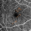
Common features of OCTA in non-proliferative diabetic retinopathy. (A) microaneurysms, (B) capillary non-perfusion area, (C) slightly enlarged foveal avascular zone, (D) abnormal vascular loops.
Figure 7
Figure 7
Optical coherence tomography angiography (OCTA) images present the foveal avascular zone, macular capillary nonperfusion and vessel density in diabetic patients. Black and white scans (A, C, and E) represent OCTA angiograms. Color map scans (B, D, and F) represent color-coded vessel density in the corresponding OCTA angiograms. With worsening diabetic retinopathy severity level, the foveal avascular zone diameters increase, and the non-perfusion area and the vessel density decrease in these images. (A, B), No diabetic retinopathy. (C, D), nonproliferative diabetic retinopathy (NPDR). (E, F), Proliferative diabetic retinopathy (PDR).

References

    1. Rathmann W, Giani G. Global prevalence of diabetes: Estimates for the year 2000 and projections for 2030. Diabetes Care (2004) 27(10):2568–9. doi: 10.2337/diacare.27.10.2568 - DOI - PubMed
    1. International diabetes federation. IDF diabetes atlas, 10th edn (2021). Brussels, Belgium. Available at: https://www.diabetesatlas.org (Accessed November 14, 2022).
    1. Yau JW, Rogers SL, Kawasaki R, Lamoureux EL, Kowalski JW, Bek T, et al. . Global prevalence and major risk factors of diabetic retinopathy. Diabetes Care (2012) 35(3):556–64. doi: 10.2337/dc11-1909 - DOI - PMC - PubMed
    1. Ruta LM, Magliano DJ, Lemesurier R, Taylor HR, Zimmet PZ, Shaw JE. Prevalence of diabetic retinopathy in type 2 diabetes in developing and developed countries. Diabetes Med (2013) 30(4):387–98. doi: 10.1111/dme.12119 - DOI - PubMed
    1. Cheung N, Mitchell P, Wong TY. Diabetic retinopathy. Lancet (2010) 376(9735):124–36. doi: 10.1016/s0140-6736(09)62124-3 - DOI - PubMed

Publication types