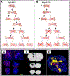
In Vivo Cellular Magnetic Imaging: Labeled vs. Unlabeled Cells
- PMID: 36589903
- PMCID: PMC9798832
- DOI: 10.1002/adfm.202207626
In Vivo Cellular Magnetic Imaging: Labeled vs. Unlabeled Cells
Abstract
Superparamagnetic iron oxide (SPIO)-labeling of cells has been applied for magnetic resonance imaging (MRI) cell tracking for over 30 years, having resulted in a dozen or so clinical trials. SPIO nanoparticles are biodegradable and can be broken down into elemental iron, and hence the tolerance of cells to magnetic labeling has been overall high. Over the years, however, single reports have accumulated demonstrating that the proliferation, migration, adhesion and differentiation of magnetically labeled cells may differ from unlabeled cells, with inhibition of chondrocytic differentiation of labeled human mesenchymal stem cells (hMSCs) as a notable example. This historical perspective provides an overview of some of the drawbacks that can be encountered with magnetic labeling. Now that magnetic particle imaging (MPI) cell tracking is emerging as a new in vivo cellular imaging modality, there has been a renaissance in the formulation of SPIO nanoparticles this time optimized for MPI. Lessons learned from the occasional past pitfalls encountered with SPIO-labeling of cells for MRI may expedite possible future clinical translation of (combined) MRI/MPI cell tracking.
Keywords: Magnetic resonance imaging; cell tracking; immune cells; magnetic particle imaging; stem cells; superparamagnetic iron oxide nanoparticles.
Conflict of interest statement
Conflict of interest statement J.W.M.B. is a paid consultant to NovaDip Biosciences SA and SuperBranche. These arrangements have been reviewed and approved by the Johns Hopkins University in accordance with its conflict-of-interest policies. C.W. and A.S-Z. have nothing to disclose.
Figures


Similar articles
-
Functional investigations on human mesenchymal stem cells exposed to magnetic fields and labeled with clinically approved iron nanoparticles.BMC Cell Biol. 2010 Apr 6;11:22. doi: 10.1186/1471-2121-11-22. BMC Cell Biol. 2010. PMID: 20370915 Free PMC article.
-
N-Alkyl-polyethylenimine 2 kDa–stabilized superparamagnetic iron oxide nanoparticles for MRI cell tracking.2011 Jan 13 [updated 2011 Feb 22]. In: Molecular Imaging and Contrast Agent Database (MICAD) [Internet]. Bethesda (MD): National Center for Biotechnology Information (US); 2004–2013. 2011 Jan 13 [updated 2011 Feb 22]. In: Molecular Imaging and Contrast Agent Database (MICAD) [Internet]. Bethesda (MD): National Center for Biotechnology Information (US); 2004–2013. PMID: 21370511 Free Books & Documents. Review.
-
In vivo tracking of adenoviral-transduced iron oxide-labeled bone marrow-derived dendritic cells using magnetic particle imaging.Eur Radiol Exp. 2023 Aug 15;7(1):42. doi: 10.1186/s41747-023-00359-4. Eur Radiol Exp. 2023. PMID: 37580614 Free PMC article.
-
Quantitative "Hot Spot" Imaging of Transplanted Stem Cells using Superparamagnetic Tracers and Magnetic Particle Imaging (MPI).Tomography. 2015 Dec;1(2):91-97. doi: 10.18383/j.tom.2015.00172. Tomography. 2015. PMID: 26740972 Free PMC article.
-
Superparamagnetic iron oxides as MPI tracers: A primer and review of early applications.Adv Drug Deliv Rev. 2019 Jan 1;138:293-301. doi: 10.1016/j.addr.2018.12.007. Epub 2018 Dec 13. Adv Drug Deliv Rev. 2019. PMID: 30552918 Free PMC article. Review.
Cited by
-
Imaging-guided precision hyperthermia with magnetic nanoparticles.Nat Rev Bioeng. 2025 Mar;3(3):245-260. doi: 10.1038/s44222-024-00257-3. Epub 2024 Nov 7. Nat Rev Bioeng. 2025. PMID: 40260131 Free PMC article.
-
Gas Vesicle-Assisted Ultrasound Imaging for Effective Anti-Tumour CAR-T Cell Immunotherapy Efficacy in Mice Model.Int J Nanomedicine. 2025 Apr 16;20:4849-4862. doi: 10.2147/IJN.S508846. eCollection 2025. Int J Nanomedicine. 2025. PMID: 40259914 Free PMC article.
-
Recent advances in non-invasive in vivo tracking of cell-based cancer immunotherapies.Biomater Sci. 2025 Apr 8;13(8):1939-1959. doi: 10.1039/d4bm01677g. Biomater Sci. 2025. PMID: 40099377 Review.
-
The role of tumor model in magnetic targeting of magnetosomes and ultramagnetic liposomes.Sci Rep. 2023 Feb 8;13(1):2278. doi: 10.1038/s41598-023-28914-4. Sci Rep. 2023. PMID: 36755030 Free PMC article.
-
Spillover can limit accurate signal quantification in MPI.Npj Imaging. 2025 May 6;3(1):20. doi: 10.1038/s44303-025-00084-0. Npj Imaging. 2025. PMID: 40604257 Free PMC article.
References
-
- Rosenberg SA, Lotze MT, Muul LM, Chang AE, Avis FP, Leitman S, Linehan WM, Robertson CN, Lee RE, Rubin JT, et al., N Engl J Med 1987, 316, 889. - PubMed
-
- Rosenberg SA, Packard BS, Aebersold PM, Solomon D, Topalian SL, Toy ST, Simon P, Lotze MT, Yang JC, Seipp CA, et al., N Engl J Med 1988, 319, 1676. - PubMed
-
- Anguille S, Smits EL, Lion E, van Tendeloo VF, Berneman ZN, The Lancet Oncology 2014, 15, e257. - PubMed
-
- Gill S, Maus MV, Porter DL, Blood Rev 2016, 30, 157. - PubMed
Grants and funding
LinkOut - more resources
Full Text Sources
Miscellaneous
