. 2022 Nov 7;5(2):100625.
doi: 10.1016/j.jhepr.2022.100625. eCollection 2023 Feb.

Metabolic reprogramming of hepatocytes by Schistosoma mansoni eggs

Affiliations

Metabolic reprogramming of hepatocytes by Schistosoma mansoni eggs

Verena von Bülow et al. JHEP Rep. .

Abstract

Background & aims: Schistosomiasis is a parasitic infection which affects more than 200 million people globally. Schistosome eggs, but not the adult worms, are mainly responsible for schistosomiasis-specific morbidity in the liver. It is unclear if S. mansoni eggs consume host metabolites, and how this compromises the host parenchyma.

Methods: Metabolic reprogramming was analyzed by matrix-assisted laser desorption/ionization mass spectrometry imaging, liquid chromatography with high-resolution mass spectrometry, metabolite quantification, confocal laser scanning microscopy, live cell imaging, quantitative real-time PCR, western blotting, assessment of DNA damage, and immunohistology in hamster models and functional experiments in human cell lines. Major results were validated in human biopsies.

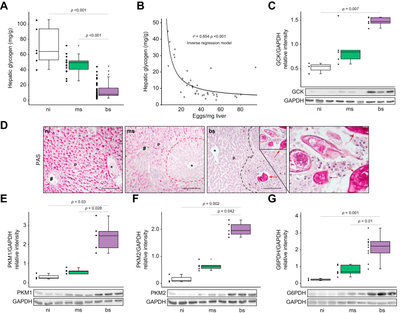
Results: The infection with S. mansoni provokes hepatic exhaustion of neutral lipids and glycogen. Furthermore, the distribution of distinct lipid species and the regulation of rate-limiting metabolic enzymes is disrupted in the liver of S. mansoni infected animals. Notably, eggs mobilize, incorporate, and store host lipids, while the associated metabolic reprogramming causes oxidative stress-induced DNA damage in hepatocytes. Administration of reactive oxygen species scavengers ameliorates these deleterious effects.

Conclusions: Our findings indicate that S. mansoni eggs completely reprogram lipid and carbohydrate metabolism via soluble factors, which results in oxidative stress-induced cell damage in the host parenchyma.

Impact and implications: The authors demonstrate that soluble egg products of the parasite S. mansoni induce hepatocellular reprogramming, causing metabolic exhaustion and a strong redox imbalance. Notably, eggs mobilize, incorporate, and store host lipids, while the metabolic reprogramming causes oxidative stress-induced DNA damage in hepatocytes, independent of the host's immune response. S. mansoni eggs take advantage of the host environment through metabolic reprogramming of hepatocytes and enterocytes. By inducing DNA damage, this neglected tropical disease might promote hepatocellular damage and thus influence international health efforts.

Keywords: DMPE, dimethyl-phosphatidylethanolamine; DNA damage; GS, glycogen synthase; GSH, reduced L-glutathione; HCC, hepatocellular carcinoma; Lipid; MALDI-MSI, matrix assisted laser desorption/ionization mass spectrometry imaging; MDA, malondialdehyde; OA, oleic acid; Oxidative stress; PAS, periodic acid-Schiff; PC, phosphatidylcholine; PDH, pyruvate dehydrogenase; PE, phosphatidylethanolamine; PLIN2, perilipin 2; Parasite; ROS, reactive oxygen species; S. japonicum, Schistosoma japonicum; S. mansoni, Schistosoma mansoni; SEA, soluble egg antigens; Schistosomiasis; TG, triglyceride; bs, bisex; flOA, fluorescently labelled OA; hRF, retention factor ∗ 100; ms, monosex; ni, non-infected.


Conflict of interest statement

B.S. and C.G.G. are consultants of TransMIT GmbH, Giessen, Germany. The other authors declare that they have no conflicts of interest. Please refer to the accompanying ICMJE disclosure forms for further details.

Figures

None
Graphical abstract
Fig. 1
Fig. 1
Infection with S. mansoni induces alterations in the composition and distribution of hepatic lipids. (A) Infection of hamsters with S. mansoni cercariae of both sexes (bs infection) or with clonal cercariae of one sex (ms infection) in order to compare egg-induced vs. worm-only effects. The distribution of lipids was analyzed by MALDI-MSI in three biological replicates of bs-infected- and ms-infected hamster, and ni (non-infected) control cryosections, respectively. (B) S. mansoni infection causes an accumulation of TGs in the granuloma area with highest levels inside the eggs (arrows). TGs were depleted in the surrounding liver tissue compared to ni controls. The panels depict the distribution of TG(16:0_18:1_18:2)+K, m/z 895.714876 in the liver of bs-infected-hamster and ni controls. (C) The distribution of lipid species differs characteristically in eggs, granuloma, and the areas surrounding granulomas. Upper left: Brightfield image. Middle left: DMPE (18:2/22:6) was found depleted in granulomas and enriched in eggs. Lower left: DMPE (18:0/22:5) was also detected with higher intensity in the eggs, but no depletion was observed for the granuloma regions. Upper right: DMPE (18:0/22:4) was mainly found in granulomas with slight enrichments in eggs. Middle right: DMPE (15:0/18:2) was found depleted in some granulomatous regions. Lower right: PC(17:2_18:3) [M+HCO2]-, m/z 810.532050, was found enriched in the outer borders of granulomas. A second set of MSI-pictures demonstrating altered lipid distribution in S. mansoni-infected hamsters is depicted in Fig. S5. These experiments were performed at least three times independently. (D) The quantification of selected lipid species revealed enhanced hepatic levels of SM and OA in bs-infected animals. These experiments were performed at least three times independently. Levels of significance are indicated in the figure (Kruskal-Wallis test). bs, bisex; Chol, cholesterol; DMPE, dimethylphosphatidylethanolamine; MALDI-MSI, matrix assisted laser desorption/ionization mass spectrometry imaging; ms, monosex; ni, non-infected; OA, oleic acid; PC, phosphatidylcholine; SM, sphingomyelin; TG, triglyceride; TO triolein.
Fig. 2
Fig. 2
S. mansoni infection exhausts the host's hepatic neutral lipid depots while parasite eggs accumulate lipids. (A) Hepatic TG content was reduced in bs-infected hamsters. (B and C) Hepatic mRNA and protein levels of fatty acid synthase (Fas) and acetyl-CoA-carboxylase 1 (Acc1), both rate-limiting enzymes for lipid synthesis, were reduced in bs-infected hamsters. Representative western blots are depicted. (D) PLIN2-staining visualized the reduction of neutral lipids in liver parenchyma (p) of bs-infected hamsters and the accumulation of neutral lipids in S. mansoni eggs (red arrows). Black arrowheads depict S. mansoni-infection-specific hepatic hemosiderin deposits, ∗central vein, #portal tract, 200x, bar 100 μm, black dotted line indicates granuloma. (E and F) Confocal microscopy clearly demonstrated the sites of lipid accumulation (white arrows) inside the eggs (liver) cultured with flOA (in green), bars 20 μm (E) and 10 μm (F). A live video showing the active uptake of flOA by an egg (liver) was deposited online (supplementary video 1) and also a 3D video demonstrating the lipid distribution inside liver-extracted eggs can be found online (supplementary video 2). (G) To prove if S. mansoni eggs mobilize and take up lipids from hepatocytes, HepG2 cells were fed with flOA, washed three times and subsequently co-cultivated with eggs in a transwell system. Around 3% of the eggs freshly isolated from the host liver took up flOA from HepG2 cells after 24 h of co-culture. The workflow of the experiment depicted schematically (left panel) and representative pictures of a liver-derived egg that took up flOA (bar 25 μm, right panel). Remarkably, nearly 100% of pre-matured in vitro-laid eggs took up flOA from HepG2 cells. These experiments were performed at least three times independently. (H) CLSM-based quantification of fluorescence intensity of individual eggs from coculture performed at 4°C (white bar) or with 4% PFA fixed HepG2 cells (purple bar) in comparison to the conventional experiment as a control (con, green bar). At least 10 eggs per condition were analyzed in each of two independent experiments. Levels of significance are indicated in the figure (Kruskal-Wallis-test). ACC, acetyl-CoA carboxylase; bs, bisex; FAS, fatty acid synthase; flOA, fluorescently labeled oleic acid; GAPDH, glyceraldehyde 3-phosphate dehydrogenase; ms, monosex; ni, non-infected; PLIN, perilipin; RFI, relative fluorescence intensity; TG, triglyceride.
Fig. 3
Fig. 3
S. mansoni infection exhausts the host's hepatic carbohydrate storage. (A) The hepatic glycogen content was diminished approximately 6-fold in livers of bs-infected hamsters. (B) Hepatic glycogen content inversely correlated with the number of eggs per mg of liver tissue. (C) Glucokinase, the first rate-limiting enzyme of glycolysis, was upregulated in livers of bs-infected hamsters. A representative western blot is shown. (D) PAS staining of histologic liver sections revealed homogenous hepatic glycogen storage in ni animals (left panel), zoned glycogen storage in livers of ms-infected hamsters with a reduction of glycogen deposits around the central veins∗ (dotted red line indicates zonation), as well as a complete absence of glycogen in the liver parenchyma (p) of bs-infected hamsters, while eggs were strongly positive stained (red arrows). ∗central vein, #portal tract, 200 x, bar 100 μm, black dotted line indicates granuloma. (E and F) Western blot analysis demonstrated the induction of rate-limiting enzymes of glycolysis like PKM1 (E) and PKM2 (F) in livers of bs-infected hamsters. (G) G6PDH, the rate-limiting enzyme of the PPP of glycolysis, was also upregulated in livers of bs-infected hamsters, n = 3-5. These experiments were performed at least three times independently. Levels of significance are indicated in the figure (Kruskal-Wallis-test and non-linear regression). bs, bisex; flOA, fluorescently labeled oleic acid; G6PDH, Glucose-6-phosphate dehydrogenase; GCK; glucokinase; ms, monosex; ni, non-infected; PAS, periodic acid-Schiff; PKM, pyruvate kinase muscle isozyme; PPP, pentose phosphate pathway.
Fig. 4
Fig. 4
S. mansoni egg-induced hepatocellular oxidative stress. (A) Hepatic MDA levels were increased in hamster livers upon bs infection. (B) SEA stimulation induced MDA in HepG2 cells. The induction was abolished by the addition of reduction equivalents in form of GSH (n = 6). (C and D) Hepatic catalase expression is reduced in hamster liver of the bs group (C, mRNA, D, protein level). (E) Hepatic catalase mRNA levels and the number of eggs per mg of liver tissue inversely correlated with exponential trend (regression analysis). Data were normalized to the control group. (F) Pyruvate quantification demonstrated SEA-induced glycolysis in HepG2 cells, which was reduced to unstimulated levels by the addition of reduction equivalents in the form of GSH. The experiment was repeated three times. Data were normalized to the control group. Kruskall-Wallis test was performed to assess group differences. These experiments were performed at least three times independently. Levels of significance are indicated in the figure (Kruskal-Wallis test). bs, bisex; CAT, catalase; Con, control; GSH, reduced L-glutathione; MDA, malondialdehyde; ms, monosex; ni, non-infected; SEA, soluble egg antigens.
Fig. 5
Fig. 5
S. mansoni eggs induce malignant signaling and oxidative stress-dependent DNA damage in HepG2 cells. (A-C) Western blot analyses revealed the activation of ERK, c-JUN, and STAT3 in SEA-stimulated HepG2 cells. The addition of GSH diminished SEA-induced HCC-associated signaling to control levels. Representative western blots are depicted. (D) γH2A.x, a marker for DNA double strand breaks, is induced in the bs group. (E) Stimulation with SEA induced γH2A.x, while the addition of GSH abolished this effect. (F) Comet assay demonstrated that SEA induced DNA damage in HepG2 cells, and that DNA damage was reduced by addition of GSH. Kruskall-Wallis test was performed to assess group differences. These experiments were performed at least three times independently. Levels of significance are indicated in the figure (Kruskal-Wallis test). bs, bisex; Con, control; γ-H2A.x, phosphorylated form of H2A histone family member X; GSH, reduced L-glutathione; ms, monosex; ni, non-infected; SEA, soluble egg antigens.
Fig. 6
Fig. 6
Validation of metabolic implications with tissue samples of a patient infected with S. mansoni. (A) Histologic specimen of a colon biopsy from a 23-year-old patient with schistosomiasis was stained for PLIN2. The signal for PLIN2 indicated an accumulation of neutral lipids inside the eggs (red arrows). (B) The positive PAS reaction demonstrated an accumulation of glycogen in most of the eggs passing the bowel wall (purple arrows). (C) Enhanced staining for PKM2 (red arrows) in granulomatous infiltrates around the eggs (black arrows). Representative stainings are depicted, bars 100 (right) and 200 μm (left). PAS, periodic acid-Schiff; PKM, pyruvate kinase muscle isozyme; PLIN, perilipin.

References

    1. WHO: Fact-Sheets Schistosomiasis 2021; Available from: https://www.who.int/news-room/fact-sheets/detail/schistosomiasis.
    1. Boissier J., Grech-Angelini S., Webster B.L., Allienne J.-F., Huyse T., Mas-Coma S., et al. Outbreak of urogenital schistosomiasis in Corsica (France): an epidemiological case study. Lancet Infect Dis. 2016;16(8):971–979. - PubMed
    1. Costain A.H., MacDonald A.S., Smits H.H. Schistosome egg migration: mechanisms, pathogenesis and host immune responses. Front Immunol. 2018;9:3042. - PMC - PubMed
    1. Schwartz C., Fallon P.G. Schistosoma "eggs-iting" the host: granuloma formation and egg excretion. Front Immunol. 2018;9:2492. - PMC - PubMed
    1. Olveda D.U., Olveda R.M., McManus D.P., Cai P., Chau T.N., Lam A.K., et al. The chronic enteropathogenic disease schistosomiasis. Int J Inf Dis. 2014;28:193–203. - PubMed