Use of radiographic and histologic scores to evaluate cats with idiopathic megacolon grouped based on the duration of their clinical signs
- PMID: 36590806
- PMCID: PMC9800827
- DOI: 10.3389/fvets.2022.1033090
Use of radiographic and histologic scores to evaluate cats with idiopathic megacolon grouped based on the duration of their clinical signs
Abstract
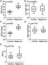
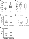
Since the duration of clinical signs could be used to identify cases of chronic constipation, in addition, prolonged duration is often associated with irreversible changes. Thus, the main objective of this study was to determine whether the duration of clinical signs of idiopathic megacolon in cats affected their diagnosis and prognosis after treatment. Medical records of cats that either had confirmed megacolon for an unknown cause (cat patients) or with normal bowels (control cats) were reviewed. Cat patients were grouped based on the duration of their clinical signs (constipation/obstipation) to cats <6 months and ≥6 months. For all feline patients, abdominal radiographs (for colonic indexes) and resected colon specimens (for histology) were assessed vs. control cats. Treatment applied to cat patients was also evaluated. Cat patients were older (p = 0.0138) and had a higher maximum colon diameter (MCD; mean 41.25 vs. 21.67 mm, p < 0.0001) and MCD/L5L ratio (1.77 vs. 0.98, p < 0.0001) than controls. Compared to cats with <6 months, cats ≥6 months showed a higher MCD (43.78 vs. 37.12 mm, p < 0.0001) and MCD/L5L ratio (1.98 vs. 1.67, p < 0.0001). Histologically, increased thickness of the smooth muscularis mucosa (54.1 vs. 22.33 μm, p < 0.05), and inner circular (743.65 vs. 482.67 μm, p < 0.05) and outer longitudinal (570.68 vs. 330.33 μm, p < 0.05) smooth muscular layers of the muscularis externa was noted only in cat patients with ≥6 months compared to controls. Similarly, fewer ganglion cells (0.93 vs. 2.87, p < 0.005) and more necrotized myocytes (2.25 vs. 0.07, p < 0.005) were observed in cats with ≥6 months. In contrast to <6 months, the majority of cats (94.4%) with ≥6 months duration did not show any response to medical treatment and therefore underwent surgery with favorable results. In conclusion, this study suggests that the duration of clinical signs should be considered in conjunction with maximal colon scores to evaluate cats for idiopathic megacolon and determine the level of treatment. Functional abnormalities of the colonic smooth muscles may be a possible cause of idiopathic megacolon in cats.
Keywords: cats; histopathology; idiopathic megacolon; radiography; signs duration.
Copyright © 2022 Abdelbaset-Ismail, Ibrahim, Sobh, Ahmed, Al-Saeed, Al-Doaiss, Al Syaad, Elmezyen and Abd-Elmaboud.
Conflict of interest statement
The authors declare that the research was conducted in the absence of any commercial or financial relationships that could be construed as a potential conflict of interest.
Figures


Similar articles
-
Alterations in colonic smooth muscle function in cats with idiopathic megacolon.Am J Vet Res. 1996 Apr;57(4):580-7. Am J Vet Res. 1996. PMID: 8712528
-
Pathogenesis, diagnosis, and therapy of feline idiopathic megacolon.Vet Clin North Am Small Anim Pract. 1999 Mar;29(2):589-603. Vet Clin North Am Small Anim Pract. 1999. PMID: 10202804 Review.
-
Radiographic diameter of the colon in normal and constipated cats and in cats with megacolon.Vet Radiol Ultrasound. 2011 Sep-Oct;52(5):516-20. doi: 10.1111/j.1740-8261.2011.01830.x. Epub 2011 May 20. Vet Radiol Ultrasound. 2011. PMID: 21599794
-
Effects of a probiotic (SLAB51™) on clinical and histologic variables and microbiota of cats with chronic constipation/megacolon: a pilot study.Benef Microbes. 2018 Jan 29;9(1):101-110. doi: 10.3920/BM2017.0023. Epub 2017 Oct 25. Benef Microbes. 2018. PMID: 29065705
-
Megacolon in cats. The role of colectomy.Vet Clin North Am Small Anim Pract. 1993 May;23(3):587-94. doi: 10.1016/s0195-5616(93)50307-8. Vet Clin North Am Small Anim Pract. 1993. PMID: 8503161 Review.
References
-
- Rosin E, Walshaw R, Mehlhaff C, Matthiesen D, Orsher R, Kusba J. Subtotal colectomy for treatment of chronic constipation associated with idiopathic megacolon in cats: 38 cases (1979-1985). J Am Vet Med Assoc. (1988) 193:850–3. - PubMed
-
- Williams JM. Colon. In:Tobias KM, Johnston SA, editors. Veterinary Surgery: Small Animal. Saint Louis, MO: Elsevier/Saunders; (2012), p. 1542–61.
LinkOut - more resources
Full Text Sources
Miscellaneous

