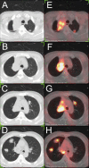
Case report: Rare epithelioid hemangioendothelioma occurs in both main bronchus and lung
- PMID: 36590968
- PMCID: PMC9799331
- DOI: 10.3389/fmed.2022.1066870
Case report: Rare epithelioid hemangioendothelioma occurs in both main bronchus and lung
Abstract
Pulmonary epithelioid hemangioendothelioma (PEH) is a rare vascular tumor of endothelial origin with low- to intermediate-grade malignant potentials. Since there is no characteristic clinical or biological marker available for PEH, most cases require a surgical lung biopsy for diagnosis. To date, although some patients with PEH reported in the literature were diagnosed through bronchoscopic biopsy, most of the patients still underwent surgical lung biopsy for confirmation. In this case report, we present a rare case diagnosed as PEH through endobronchial biopsies due to the presence of an intraluminal mass that blocked the trachea and caused atelectasis in the right upper lobe. Moreover, since surgery was not appropriate for this patient with unresectable bilateral multiple nodules, we adopted genetic analysis using NGS to provide a guide for personalized treatment. Then, based on the NGS results, the patient was treated with anti-PD-1 mAb and sirolimus for 1 year and has been stable in a 1-year follow-up examination.
Keywords: POLE (P286R) mutation; bronchoscopic; case report; genetic analysis; pulmonary epithelioid hemangioendothelioma.
Copyright © 2022 Gong, Tian, Wang, Mu, Geng, Hao, Zhong, Zhang, Jiang, Wang and Bao.
Conflict of interest statement
The authors declare that the research was conducted in the absence of any commercial or financial relationships that could be construed as a potential conflict of interest.
Figures


Similar articles
-
Clinico-radiological features and next generation sequencing of pulmonary epithelioid hemangioendothelioma: A case report and review of literature.Thorac Cancer. 2017 Nov;8(6):687-692. doi: 10.1111/1759-7714.12474. Epub 2017 Aug 4. Thorac Cancer. 2017. PMID: 28777494 Free PMC article. Review.
-
Pulmonary Epithelioid Hemangioendothelioma Diagnosed With Endobronchial Biopsies: A Case Report and Literature Review.J Bronchology Interv Pulmonol. 2016 Apr;23(2):168-73. doi: 10.1097/LBR.0000000000000230. J Bronchology Interv Pulmonol. 2016. PMID: 27058721 Review.
-
[Case report of pulmonary epithelioid hemangioendothelioma].Nihon Kokyuki Gakkai Zasshi. 2003 Feb;41(2):144-9. Nihon Kokyuki Gakkai Zasshi. 2003. PMID: 12722336 Review. Japanese.
-
Malignant pulmonary epithelioid hemangioendothelioma with hilar lymph node metastasis.Ann Diagn Pathol. 2011 Jun;15(3):207-12. doi: 10.1016/j.anndiagpath.2010.02.012. Epub 2010 Jun 16. Ann Diagn Pathol. 2011. PMID: 20952289
-
A Rare Case of Pulmonary Epithelioid Hemangioendothelioma Presenting with Skin Metastasis.Arch Plast Surg. 2016 May;43(3):284-7. doi: 10.5999/aps.2016.43.3.284. Epub 2016 May 18. Arch Plast Surg. 2016. PMID: 27218028 Free PMC article.
Cited by
-
Epithelioid hemangioendothelioma of the retroperitoneal giant type treated with Toripalimab: A case report.Front Immunol. 2023 Mar 17;14:1116944. doi: 10.3389/fimmu.2023.1116944. eCollection 2023. Front Immunol. 2023. PMID: 37006308 Free PMC article.
References
-
- PáLföldi R, Radács M, Csada E, Molnár Z, Pintér S, Tiszlavicz L, et al. Pulmonary epithelioid haemangioendothelioma studies in vitro and in vivo: new diagnostic and treatment methods. In Vivo. (2013) 27:221–5. - PubMed
-
- Liu H, Wang J, Lang J, Zhang X. Pulmonary epithelioid hemangioendothelioma: imaging and clinical features. J Compu Assist Tomogr. (2021) 45:788–94. - PubMed
Publication types
LinkOut - more resources
Full Text Sources

