

Decellularized matrix for repairing intervertebral disc degeneration: Fabrication methods, applications and animal models
- PMID: 36590980
- PMCID: PMC9800636
- DOI: 10.1016/j.mtbio.2022.100523
Decellularized matrix for repairing intervertebral disc degeneration: Fabrication methods, applications and animal models
Abstract
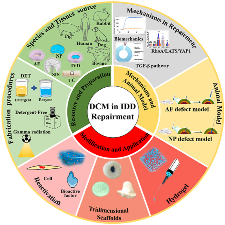
Intervertebral disc degeneration (IDD)-induced low back pain significantly influences the quality of life, placing a burden on public health systems worldwide. Currently available therapeutic strategies, such as conservative or operative treatment, cannot effectively restore intervertebral disc (IVD) function. Decellularized matrix (DCM) is a tissue-engineered biomaterial fabricated using physical, chemical, and enzymatic technologies to eliminate cells and antigens. By contrast, the extracellular matrix (ECM), including collagen and glycosaminoglycans, which are well retained, have been extensively studied in IVD regeneration. DCM inherits the native architecture and specific-differentiation induction ability of IVD and has demonstrated effectiveness in IVD regeneration in vitro and in vivo. Moreover, significant improvements have been achieved in the preparation process, mechanistic insights, and application of DCM for IDD repair. Herein, we comprehensively summarize and provide an overview of the roles and applications of DCM for IDD repair based on the existing evidence to shed a novel light on the clinical treatment of IDD.
Keywords: (3D), three-dimensional; (AF), annular fibers; (AFSC), AF stem cells; (APNP), acellular hydrogel descendent from porcine NP; (DAF-G), decellularized AF hydrogel; (DAPI), 4,6-diamidino-2-phenylindole; (DCM), decellularized matrix; (DET), detergent-enzymatic treatment; (DWJM), Wharton's jelly matrix; (ECM), extracellular matrix; (EVs), extracellular vesicles; (Exos), exosome; (IDD), intervertebral disc degeneration; (IVD), intervertebral disc; (LBP), Low back pain; (NP), nucleus pulposus; (NPCS), NP-based cell delivery system; (PEGDA/DAFM), polyethylene glycol diacrylate/decellularized AF matrix; (SD), sodium deoxycholate; (SDS), sodium dodecyl sulfate; (SIS), small intestinal submucosa; (TGF), transforming growth factor; (bFGF), basic fibroblast growth factor; (hADSCs), human adipose-derived stem cells; (hDF), human dermal fibroblast; (iAF), inner annular fibers; (oAF), outer annular fibers; (sGAG), sulfated glycosaminoglycan; Decellularized matrix; Intervertebral disc degeneration; Regenerative medicine; Tissue engineering.
© 2022 The Authors.
Conflict of interest statement
The authors declare that they have no known competing financial interests or personal relationships that could have appeared to influence the work reported in this paper.
Figures

Similar articles
-
Extracellular Matrix From Decellularized Wharton's Jelly Improves the Behavior of Cells From Degenerated Intervertebral Disc.Front Bioeng Biotechnol. 2020 Mar 27;8:262. doi: 10.3389/fbioe.2020.00262. eCollection 2020. Front Bioeng Biotechnol. 2020. PMID: 32292779 Free PMC article.
-
Decellularized Disc Hydrogels for hBMSCs tissue-specific differentiation and tissue regeneration.Bioact Mater. 2021 Mar 22;6(10):3541-3556. doi: 10.1016/j.bioactmat.2021.03.014. eCollection 2021 Oct. Bioact Mater. 2021. PMID: 33842740 Free PMC article.
-
Injectable decellularized nucleus pulposus-based cell delivery system for differentiation of adipose-derived stem cells and nucleus pulposus regeneration.Acta Biomater. 2018 Nov;81:115-128. doi: 10.1016/j.actbio.2018.09.044. Epub 2018 Sep 27. Acta Biomater. 2018. PMID: 30267879
-
Growth and differentiation factor-5 contributes to the structural and functional maintenance of the intervertebral disc.Cell Physiol Biochem. 2015;35(1):1-16. doi: 10.1159/000369670. Epub 2015 Jan 2. Cell Physiol Biochem. 2015. PMID: 25547527 Review.
-
Research progress in decellularized extracellular matrix hydrogels for intervertebral disc degeneration.Biomater Sci. 2023 Mar 14;11(6):1981-1993. doi: 10.1039/d2bm01862d. Biomater Sci. 2023. PMID: 36734099 Review.
Cited by
-
Biomimetic Microchannel Integrated Silk Fibroin Scaffold for Regeneration of Intervertebral Disc Degeneration.Biomater Res. 2025 May 28;29:0203. doi: 10.34133/bmr.0203. eCollection 2025. Biomater Res. 2025. PMID: 40438125 Free PMC article.
-
Umbilical cord mesenchymal stem cells for regenerative treatment of intervertebral disc degeneration.Front Cell Dev Biol. 2023 Aug 3;11:1215698. doi: 10.3389/fcell.2023.1215698. eCollection 2023. Front Cell Dev Biol. 2023. PMID: 37601097 Free PMC article. Review.
-
Decellularized nucleus pulposus matrix/chitosan hybrid hydrogel combined with nucleus pulposus stem cells and GDF5-loaded microspheres for intervertebral disc degeneration prevention.Mol Med. 2024 Jan 10;30(1):7. doi: 10.1186/s10020-024-00777-z. Mol Med. 2024. PMID: 38200442 Free PMC article.
-
A novel spherical GelMA-HAMA hydrogel encapsulating APET×2 polypeptide and CFIm25-targeting sgRNA for immune microenvironment modulation and nucleus pulposus regeneration in intervertebral discs.J Nanobiotechnology. 2024 Sep 12;22(1):556. doi: 10.1186/s12951-024-02783-z. J Nanobiotechnology. 2024. PMID: 39267105 Free PMC article.
-
Engineering cell-derived extracellular matrix for peripheral nerve regeneration.Mater Today Bio. 2024 Jun 13;27:101125. doi: 10.1016/j.mtbio.2024.101125. eCollection 2024 Aug. Mater Today Bio. 2024. PMID: 38979129 Free PMC article. Review.
References
-
- Chan L.K., Leung V.Y., Tam V., Lu W.W., Sze K.Y., Cheung K.M. Decellularized bovine intervertebral disc as a natural scaffold for xenogenic cell studies. Acta Biomater. 2013;9(2):5262–5272. - PubMed
-
- Knezevic N.N., Candido K.D., Vlaeyen J.W.S., Van Zundert J., Cohen S.P. Low back pain. Lancet (London, England) 2021;398(10294):78–92. - PubMed
-
- Yu L., Sun Z.J., Tan Q.C., Wang S., Wang W.H., Yang X.Q., Ye X.J. Thermosensitive injectable decellularized nucleus pulposus hydrogel as an ideal biomaterial for nucleus pulposus regeneration. J. Biomater. Appl. 2020;35(2):182–192. - PubMed
-
- Hoy D., Brooks P., Blyth F., Buchbinder R. The Epidemiology of low back pain, Best practice & research. Clin. Rheumatol. 2010;24(6):769–781. - PubMed
-
- Benzakour T., Igoumenou V., Mavrogenis A.F., Benzakour A. Current concepts for lumbar disc herniation. Int. Orthop. 2019;43(4):841–851. - PubMed
Publication types
LinkOut - more resources
Full Text Sources
Miscellaneous
