

Trimethine Cyanine Dyes as NA-Sensitive Probes for Visualization of Cell Compartments in Fluorescence Microscopy
- PMID: 36591208
- PMCID: PMC9798395
- DOI: 10.1021/acsomega.2c05231
Trimethine Cyanine Dyes as NA-Sensitive Probes for Visualization of Cell Compartments in Fluorescence Microscopy
Abstract
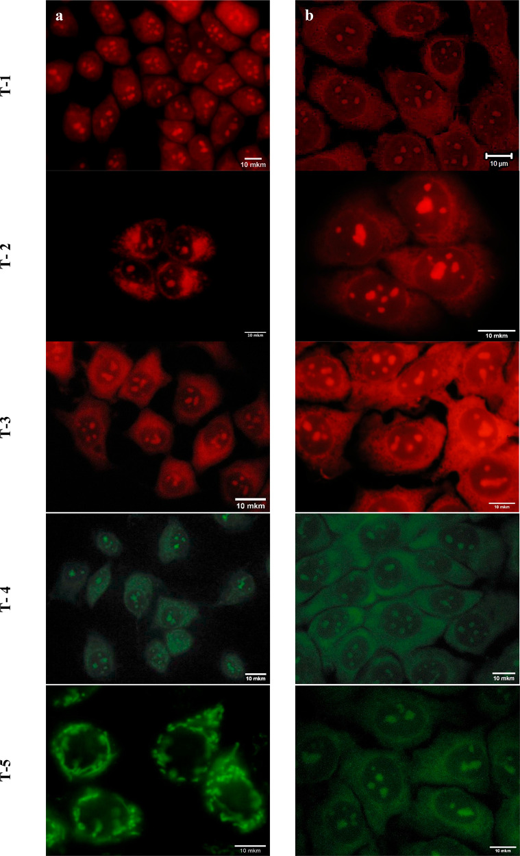
We propose symmetrical cationic trimethine cyanine dyes with β-substituents in the polymethine chain based on modified benzothiazole and benzoxazole heterocycles as probes for the detection and visualization of live and fixed cells by fluorescence microscopy. The spectral-luminescent properties of trimethine cyanines have been characterized for free dyes and in the presence of nucleic acids (NA) and globular proteins. The studied cyanines are low to moderate fluorescent when free, but in the presence of NA, they show an increase in emission intensity up to 111 times; the most pronounced emission increase was observed for the dyes T-2 in the presence of dsDNA and T-1 with RNA. Spectral methods showed the binding of all dyes to nucleic acids, and different interaction mechanisms have been proposed. The ability to visualize cell components of the studied dyes has been evaluated using different human cell lines (MCF-7, A2780, HeLa, and Hs27). We have shown that all dyes are cell-permeant staining nucleus components, probably RNA-rich nucleoli with background fluorescence in the cytoplasm, except for the dye T-5. The dye T-5 selectively stains some structures in the cytoplasm of MCF-7 and A2780 cells associated with mitochondria or lysosomes. This effect has also been confirmed for the normal type of cell line-human foreskin fibroblasts (Hs27). The costaining of dye T-5 with MitoTracker CMXRos Red demonstrates specificity to mitochondria at a concentration of 0.1 μM. Colocalization analysis has shown signals overlapping of dye T-5 and MitoTracker CMXRos Red (Pearson's Coefficient value = 0.92 ± 0.04). The photostability study shows benzoxazole dyes to be up to ∼7 times more photostable than benzothiazole ones. Moreover, studied benzoxazoles are less cytotoxic at working concentrations than benzothiazoles (67% of cell viability for T-4, T-5 compared to 12% for T-1, and ∼30% for T-2, T-3 after 24 h). Therefore, the benzoxazole T-4 dye is proposed for nucleic acid detection in vitro and intracellular fluorescence imaging of live and fixed cells. In contrast, the benzoxazole dye T-5 is proposed as a good alternative to commercial dyes for mitochondria staining in the green-yellow region of the spectrum.
© 2022 The Authors. Published by American Chemical Society.
Conflict of interest statement
The authors declare no competing financial interest.
Figures


References
-
- Hickey S. M.; Ung B.; Bader C.; Brooks R.; Lazniewska J.; Johnson I. R. D.; Sorvina A.; Logan J.; Martini C.; Moore C. R.; Karageorgos L.; Sweetman M. J.; Brooks D. A. Fluorescence Microscopy – An Outline of Hardware, Biological Handling, and Fluorophore Considerations. Cells 2022, 11 (1), 35. 10.3390/cells11010035. - DOI - PMC - PubMed
-
- Hua X.-W.; Bao Y.-W.; Zeng J.; Wu F.-G. Nucleolus-Targeted Red Emissive Carbon Dots with Polarity-Sensitive and Excitation-Independent Fluorescence Emission: High-Resolution Cell Imaging and in Vivo Tracking. ACS Appl. Mater. Interfaces. 2019, 11 (36), 32647–32658. 10.1021/acsami.9b09590. - DOI - PubMed
LinkOut - more resources
Full Text Sources
