. 2022 Dec 14:12:986343.
doi: 10.3389/fonc.2022.986343. eCollection 2022.

Plasma exosomal miR-320d, miR-4479, and miR-6763-5p as diagnostic biomarkers in epithelial ovarian cancer

Affiliations

Plasma exosomal miR-320d, miR-4479, and miR-6763-5p as diagnostic biomarkers in epithelial ovarian cancer

Shiwen Wang et al. Front Oncol. .

Abstract

Background: Exosomal miRNA had been proved as the promising biomarkers for multiple cancers including epithelial ovarian cancer (EOC). This study aimed to validate the diagnostic accuracy of exosomal miR-320d, miR-4479, and miR-6763-5p for EOC.

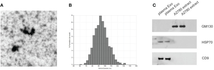
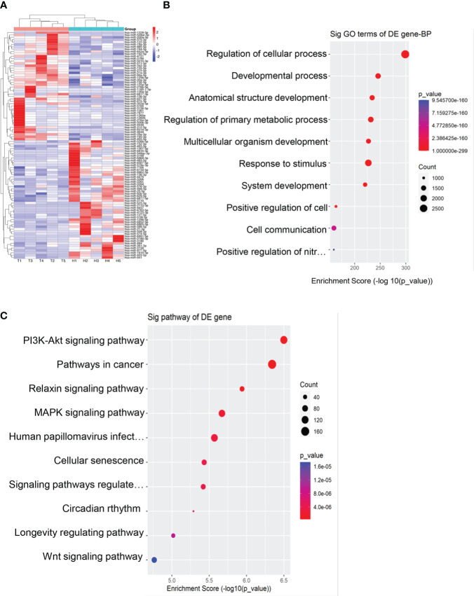
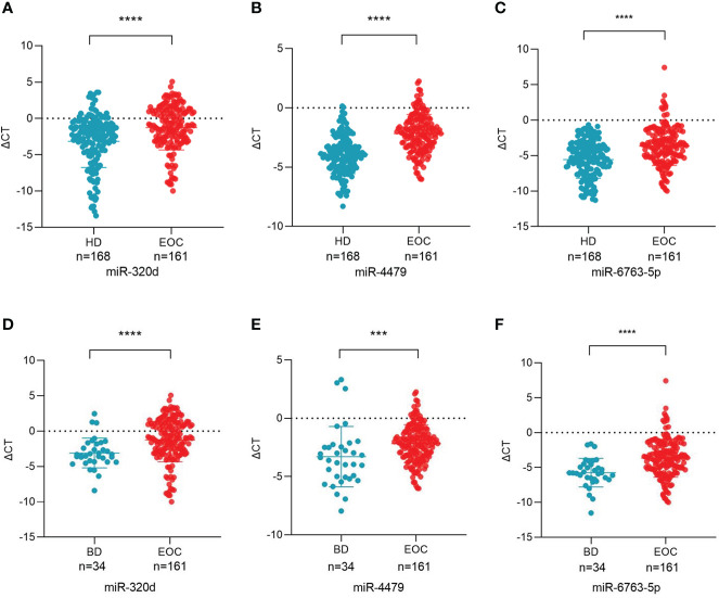
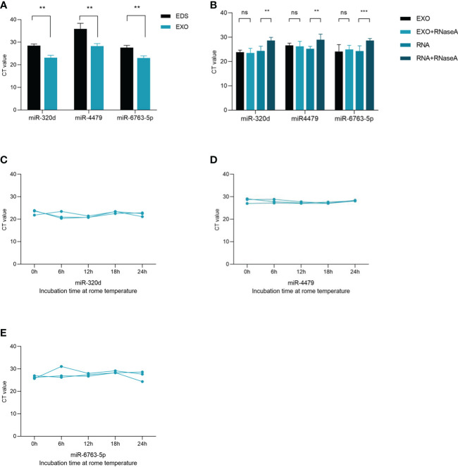
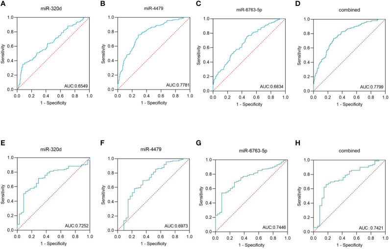
Materials and methods: Exosomes isolated from the plasma by ultracentrifugation were verified using TEM, qNano and western blot. MiRNAs sequencing was used to screen out the differential exosomal miRNAs and miR-320d, miR-4479, and miR-6763-5p were selected as candidates, which were further verified by RT-qPCR in 168 healthy donors and 161 primary EOC patients. Besides, the diagnostic accuracy of these three exosomal miRNAs were evaluated using the receiver operating characteristic curve (ROC).

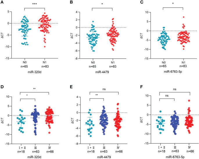
Results: MiRNAs sequencing revealed 95 differential exosomal miRNAs between EOC patients and healthy donors. Subsequently, exosomal miR-320d, miR-4479, and miR-6763-5p were significantly down regulated in EOC patients compared with healthy controls and benign patients. More importantly, these three miRNAs could serve as circulating diagnostics biomarkers for EOC, possessing areas under the curve (AUC) of 0.6549, 0.7781, and 0.6834, respectively. Moreover, these three exosomal miRNAs levels were closely associated with lymph node metastasis, meanwhile exosomal miR-320d and miR-4479 expression was related to tumor stage.

Conclusion: Exosomal miR-320d, miR-4479, and miR-6763-5p might serve as potential biomarkers for EOC.

Keywords: biomarkers; diagnosis; epithelial ovarian cancer; exosomes; miRNAs.


Conflict of interest statement

The authors declare that the research was conducted in the absence of any commercial or financial relationships that could be construed as a potential conflict of interest.

Figures

Figure 1
Figure 1
Identification of isolated exosomes. (A) TEM image showed the data of exosomes from EOC plasma samples with 50–150 nm diameter. (B) Distribution of exosomes with 50–150 nm diameter; the samples were obtained from EOC plasma samples based on the qNano system. (C) HSP70, CD9, and GM130 as common exosome markers, were analyzed by western blot.
Figure 2
Figure 2
Exosomal miRNAs profile of the EOC patients. (A) A heat map was generated after supervised hierarchical cluster analysis. The differential exosomal miRNAs expression is shown in red (upregulation) or blue (downregulation) (P < 0.05). (B). Candidate target gene GO enrichment analysis. (C) Candidate target gene KEGG enrichment analysis.
Figure 3
Figure 3
Plasma exosomal miR-320d, miR-4479, and miR-6763-5p are potential biomarkers of EOC. (A-C) Scatter plots comparing the exosomal miR-320d, miR-4479, and miR-6763-5p levels in the plasma of healthy donors (n = 168) and EOC patients (n = 161) (****P < 0.0001). (D-F) Scatter plots comparing the exosomal miR-320d, miR-4479, and miR-6763-5p levels in the plasma of EOC patients (n = 161) and benign diseases patients (n=34) (***P < 0.001, ****P < 0.0001). HD, Healthy donor; EOC, epithelial ovarian cancer; BD, benign diseases.
Figure 4
Figure 4
Characterization of identified three plasma exosomal miRNAs. (A) Expression levels of miR-320d, miR-4479, and miR-6763-5p from plasma exosomes (EXO) and exosome-depleted supernatant (EDS). (B) Expression levels of both miRNAs in exosomes treated with RNase A or in isolated RNA. (C–E) The expressions of three plasma exosomal miRNAs when incubated at room temperature (**P < 0.01, ***P < 0.001, ns, not significant).
Figure 5
Figure 5
Diagnostic role of plasma exosomal miRNAs expression levels in EOC patients. The AUCs of miR-320d (A), miR-4479 (B), miR-6763-5p (C), and both (D) in EOC patients relative to healthy donors. The AUCs of miR-320d (E), miR-4479 (F), miR-6763-5p (G) and both (H) in EOC patients relative to benign diseases patients. AUC, Areas under the curve.
Figure 6
Figure 6
Association between the plasma exosomal miRNAs and the clinical data of EOC patients. (A-C) The statistical association between the levels of exosomal miR-320d, miR-4479, and miR-6763-5p and lymph node metastasis. (D-F) The plasma exosomal miR-320d, miR-4479, and miR-6763-5p expression in the FIGO stage. (*P <0.05, **P < 0.01, ***P < 0.001, ns: not significant). HD, Healthy donor; EOC, Epithelial ovarian cancer.

References

    1. Matulonis UA, Sood AK, Fallowfield L, Howitt BE, Sehouli J, Karlan BY. Ovarian cancer. Nat Rev Dis Primers (2016) 2:16061. doi: 10.1038/nrdp.2016.61 - DOI - PMC - PubMed
    1. Lheureux S, Gourley C, Vergote I, Oza AM. Epithelial ovarian cancer. Lancet (London England) (2019) 393(10177):1240–53. doi: 10.1016/S0140-6736(18)32552-2 - DOI - PubMed
    1. Sung H, Ferlay J, Siegel RL, Laversanne M, Soerjomataram I, Jemal A, et al. . Global cancer statistics 2020: GLOBOCAN estimates of incidence and mortality worldwide for 36 cancers in 185 countries. CA Cancer J Clin (2021) 71(3):209–49. doi: 10.3322/caac.21660 - DOI - PubMed
    1. Chen M, Zhao H. Next-generation sequencing in liquid biopsy: Cancer screening and early detection. Hum Genomics (2019) 13(1):34. doi: 10.1186/s40246-019-0220-8 - DOI - PMC - PubMed
    1. Yanez-Mo M, Siljander PR, Andreu Z, Zavec AB, Borras FE, Buzas EI, et al. . Biological properties of extracellular vesicles and their physiological functions. J Extracell Vesicles (2015) 4:27066. doi: 10.3402/jev.v4.27066 - DOI - PMC - PubMed

LinkOut - more resources