. 2023 Aug;149(9):5885-5899.
doi: 10.1007/s00432-022-04554-5. Epub 2023 Jan 2.

Involvement of angiogenesis in cancer-associated acinar-to-ductal metaplasia lesion of pancreatic cancer invasive front

Affiliations

Involvement of angiogenesis in cancer-associated acinar-to-ductal metaplasia lesion of pancreatic cancer invasive front

Shuang Fei et al. J Cancer Res Clin Oncol. 2023 Aug.

Abstract

Purpose: This study aimed to demonstrate the involvement of angiogenesis in cancer-associated acinar-to-ductal metaplasia (CA-ADM) lesion of invasive front pancreatic ductal adenocarcinoma (PDAC) and investigate the possible mechanism.

Methods: Tissue samples from 128 patients with PDAC and 36 LSL-KrasG12D/+; LSL-Trp53R172H/+; Pdx-1-Cre mice were analyzed. Immunohistochemical assay was performed using HE, anti-CK19 and anti-amylase to confirm the presence of CA-ADM lesions, using anti-CD34 and anti-CD31 to measure microvessel density (MVD), and using anti-CD68, anti-CD163, anti-iNOS, or anti-MMP9 to evaluate the immune microenvironment. We performed multiplex immunohistochemical assay to detect the co-expression of MMP9 and CD68 on macrophage. We examined clinical outcomes and other clinicopathological factors to determine the significance of high-level MVD of CA-ADM on survival and liver metastasis. We performed tube formation assay to evaluate the effect of macrophage on angiogenic capacity in vitro.

Results: Angiogenesis was significantly abundant in CA-ADM lesions compared with that in PDAC lesions in human and mouse tissues. High-level MVD in CA-ADM lesions was an independent predictor of poor prognosis (P = 0.0047) and the recurrence of liver metastasis (P = 0.0027). More CD68-positive and CD163-positive macrophages were detected in CA-ADM lesions than in PDAC. The percentage of CD68-positive macrophages was positively correlated with MVD in CA-ADM lesions. Multiplex-immunostaining revealed that MMP9 was expressed in CD68-positive macrophages of CA-ADM lesions. In CA-ADM lesions, the percentage of macrophages was positively correlated with MMP9 expression, which positively correlated with microvessel density.

Conclusion: CA-ADM related angiogenesis is a promising predictive marker for poor prognosis of PDAC and may provide an attractive therapeutic target for PDAC.

Keywords: Angiogenesis; Cancer-associated acinar-to-ductal metaplasia; Liver metastasis; Macrophages; Pancreatic cancer; Prognosis.


Conflict of interest statement

The authors declare no competing interests.

The authors have no relevant financial or non-financial interests to disclose.

Figures

Fig. 1
Fig. 1
Cancer-associated acinar-to-ductal metaplasia (CA-ADM) lesions are formed within the invasive front of pancreatic ductal adenocarcinoma (PDAC). a Human PDAC samples stained with HE. The boundaries between the tumor and the acini divide two histopathologically distinct regions, the non-invasive front and the invasive front. In the non-invasive front, the tumor is encapsulated by stromal cells. The invasive front is associated with CA-ADM lesions. b, c HE, CK19 and amylase staining in serial sections of human and mouse PDAC. Scale bars, 100 µm
Fig. 2
Fig. 2
MVD level was higher in CA-ADM lesions than in PDAC lesions in KPC model mice. a Representative micrographs of CD34 staining in CA-ADM and PDAC lesions in KPC mice. The arrowheads indicate microvessels in CA-ADM and PDAC lesions. b Microvessel counts per field in CA-ADM and PDAC lesions. CA-ADM (n = 26), PDAC (n = 36). *P < 0.05. c, d The correlation plot shows that microvessel counts per field between CA-ADM and PDAC lesions are highly correlated (n = 26). Data shown represent the mean ± SD. Scale bars, 100 µm. ****P < 0.0001
Fig. 3
Fig. 3
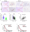
MVD level was higher in CA-ADM lesions than that in PDAC lesions in human PDAC samples. a Representative micrographs of CD31 staining in CA-ADM and PDAC lesions in human PDAC samples. The arrowheads indicate microvessels (stained with CD31) in the CA-ADM and PDAC lesion. b Microvessel counts per field in the CA-ADM lesions are higher than those in the PDAC lesions. CA-ADM (n = 120), PDAC (n = 128). ****P < 0.0001. c The comparison of microvessel counts per field between CA-ADM and PDAC lesions (n = 120). ****P < 0.0001. d The correlation plot shows that microvessel counts per field between CA-ADM and PDAC lesions are moderately correlated (n = 120). e Kaplan–Meier analysis of overall survival according to MVD in human ADM-like lesions (n = 120). High-level MVD in CA-ADM lesions is associated with shorter patient survival. f Kaplan–Meier analysis of overall survival based on MVD in human PDAC lesions (n = 128). High-level MVD of PDAC lesion is not associated with prognosis. Data shown represent the mean ± SD. Scale bars, 100 µm
Fig. 4
Fig. 4
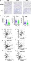
Macrophages were more frequently observed in CA-ADM lesions than in PDAC lesions. a Representative micrographs of CD68, iNOS and CD163 staining in CA-ADM and PDAC lesions of human PDAC samples. b Percentages of CD68-, CD163- and iNOS-positive cells in CA-ADM and PDAC lesions are shown in graphs. CD68, CD163 and iNOS staining was performed in CA-ADM lesions (n = 96, 96 and 79, respectively) and PDAC lesions (n = 97, 97 and 80, respectively). **P < 0.01, ****P < 0.0001. c, d The correlation plots show that the expression of CD68 and CD163 are positively correlated with the MVD in CA-ADM lesions (n = 96). e, f The correlation plots show that the expression of CD68 and CD163 are positively correlated with MVD in PDAC lesions (n = 97). g, h The correlation plots show that the expression of iNOS is not correlated with the MVD in CA-ADM or PDAC lesions (n = 79, 80 respectively). Data shown represent the mean ± SD. Scale bars, 100 µm
Fig. 5
Fig. 5
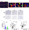
MMP9 was co-localized with CD68-positive macrophages and correlated with MVD levels in CA-ADM lesions. a Representative micrographs of CD68 and MMP9 staining via mIHC analysis in CA-ADM lesions of human PDAC samples. The arrowheads indicate co-expression areas. b Representative micrographs of CD31, CD68 and MMP9 staining in CA-ADM and PDAC lesions in human PDAC samples. c MMP9 staining in CA-ADM and PDAC lesions of human PDAC samples. CA-ADM (n = 91), PDAC (n = 92). **P < 0.01. d, e The correlation plots show that the percentage of MMP9 positive cells is correlated with the percentage of CD68 positive cells and MVD in CA-ADM lesions (n = 91). Data shown represent the mean ± SD. Scale bars, 100 µm

References

    1. Al-Abd AM, Alamoudi AJ, Abdel-Naim AB, Neamatallah TA, Ashour OM (2017) Anti-angiogenic agents for the treatment of solid tumors: potential pathways, therapy and current strategies—a review. J Adv Res 8:591–605. 10.1016/j.jare.2017.06.006 - PMC - PubMed
    1. Ansari D, Tingstedt B, Andersson B, Holmquist F, Sturesson C, Williamsson C, Sasor A, Borg D, Bauden M, Andersson R (2016) Pancreatic cancer: yesterday, today and tomorrow. Future Oncol 12:1929–1946. 10.2217/fon-2016-0010 - PubMed
    1. Ardito CM, Grüner BM, Takeuchi KK, Lubeseder-Martellato C, Teichmann N, Mazur PK, DelGiorno KE, Carpenter ES, Halbrook CJ, Hall JC (2012) EGF receptor is required for KRAS-induced pancreatic tumorigenesis. Cancer Cell 22:304–317. 10.1016/j.ccr.2012.07.024 - PMC - PubMed
    1. Augoff K, Hryniewicz-Jankowska A, Tabola R, Stach K (2022) MMP9: a tough target for targeted therapy for cancer. Cancers 14:1847. 10.3390/cancers14071847 - PMC - PubMed
    1. Brune K, Abe T, Canto M, O’Malley L, Klein AP, Maitra A, Adsay NV, Fishman EK, Cameron JL, Yeo CJ (2006) Multifocal neoplastic precursor lesions associated with lobular atrophy of the pancreas in patients having a strong family history of pancreatic cancer. Am J Surg Pathol 30:1067 - PMC - PubMed