Review
. 2023 Feb 21;82(3):250-260.
doi: 10.1093/jnen/nlac123.

MYB/MYBL1::QKI fusion-positive diffuse glioma

Affiliations
Review

MYB/MYBL1::QKI fusion-positive diffuse glioma

Ye Yoon Suh et al. J Neuropathol Exp Neurol. .

Abstract

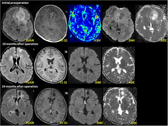
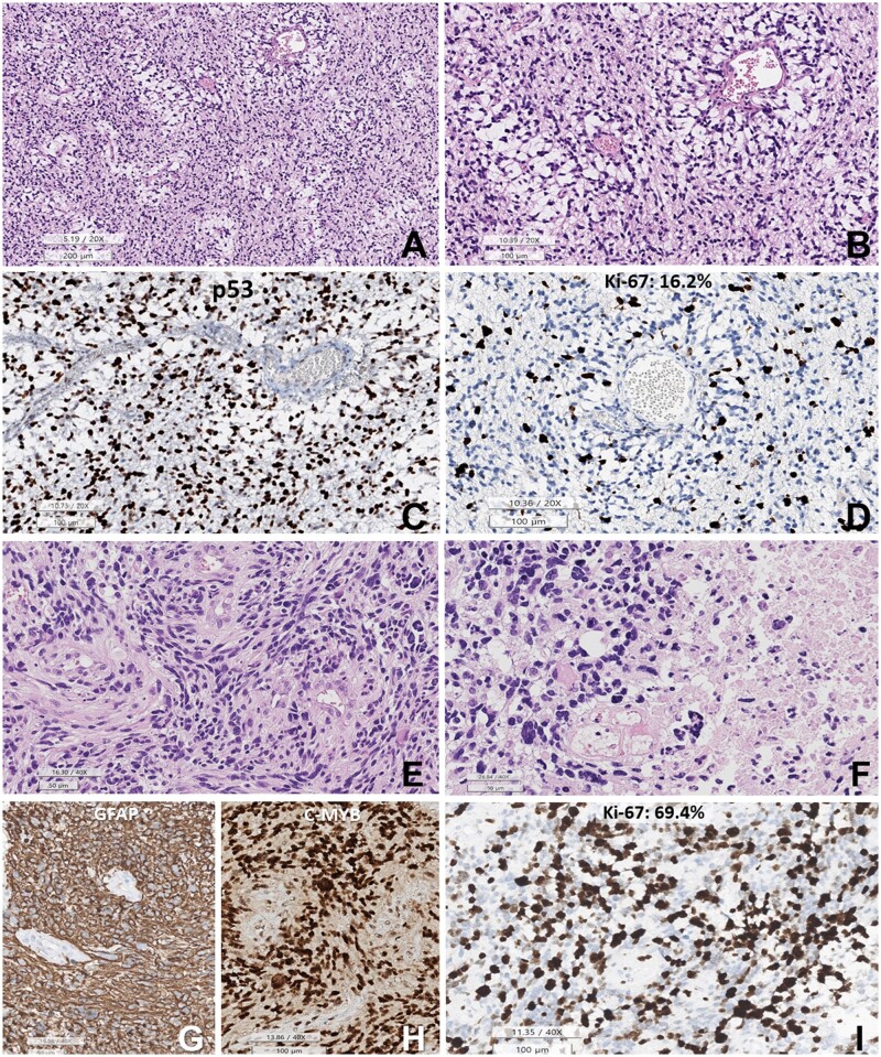
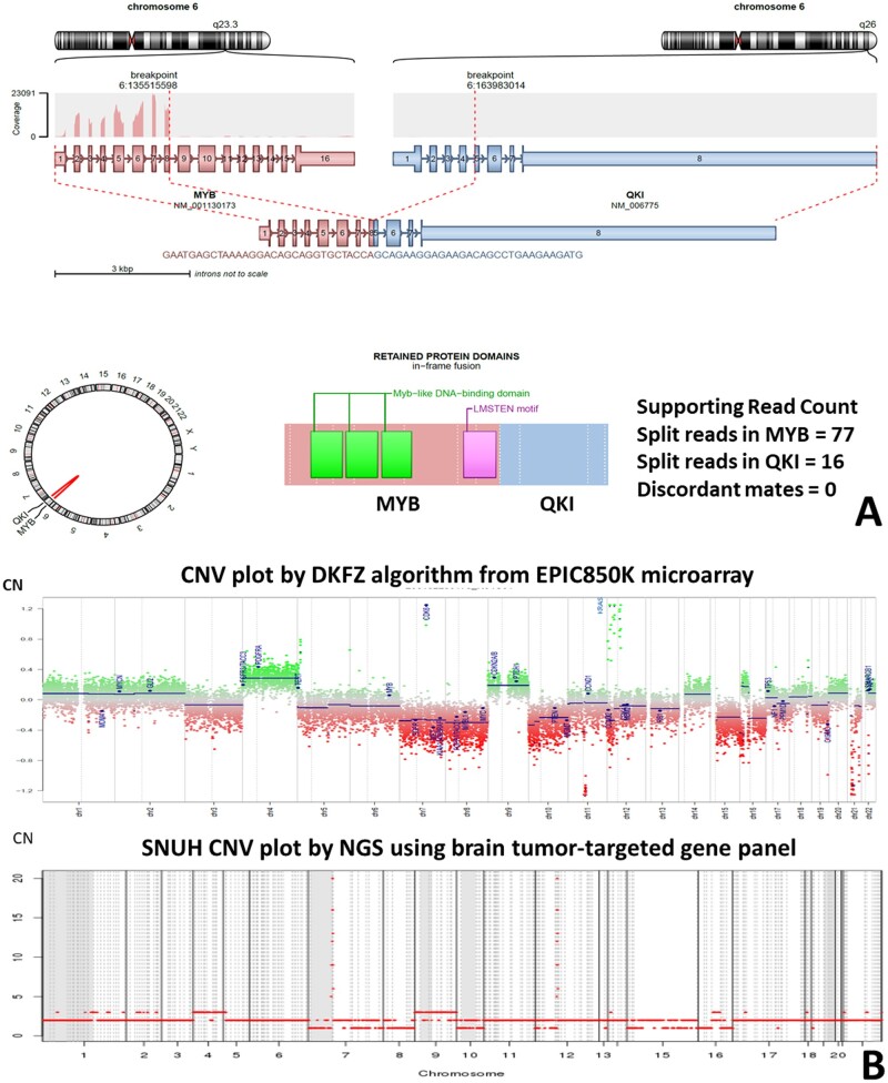
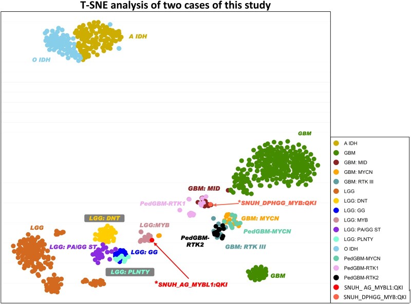
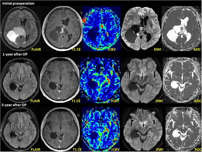
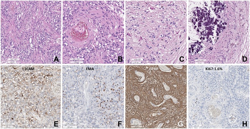
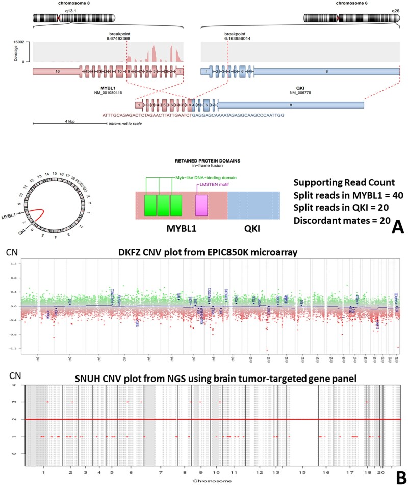
The MYB/MYBL1::QKI fusion induces the protooncogene, MYB, and deletes the tumor suppressor gene, QKI. MYB/MYBL1::QKI rearrangement was previously reported only in angiocentric glioma (AG) and diffuse low-grade glioma. This report compares 2 tumors containing the MYB/MYBL1::QKI fusion: a diffuse pediatric-type high-grade glioma (DPedHGG) in an 11-year-old boy and an AG in a 46-year-old woman. We used immunohistochemistry, next-generation sequencing, and methylation profiling to characterize each tumor and compare our findings to the literature on AG and tumors with the MYB/MYBL1::QKI rearrangement. Both tumors were astrocytic with angiocentric patterns. The MYB::QKI fusion-positive DPedHGG, which recurred once, was accompanied by TP53 mutation and amplification of CDK6 and KRAS, suggesting malignant transformation secondary to additional genetic aberrations. The second case was the adult AG with MYBL1::QKI fusion, which mimicked ependymoma based on histopathology and its dot- and ring-like epithelial membrane antigen positivity. Combined with a literature review, our results suggest that MYB/MYBL1 alterations are not limited to low-grade gliomas, including AG. AG is most common in the cerebra of children and adolescents but exceptional cases occur in adults and the acquisition of additional genetic mutations may contribute to high-grade glioma. These cases further demonstrate that molecular characteristics, morphologic features, and clinical context are essential for diagnosis.

Keywords: MYB/MYBL1-alteration; Angiocentric glioma; Methylation profile; Next-generation sequencing; Pediatric-type high-grade glioma.


Figures

Figure 1.
Figure 1.
DPedHGG with MYB::QKI fusion-positivity in a 11-year-old boy presenting with recurrent seizure. Initial MRI shows a well-defined, 3.6 cm × 3.0 cm × 2.4 cm (anteroposterior × transverse × craniocaudal) tumor in the left frontal lobe. The axial T2 FLAIR image shows high intensity with necrosis and restricted diffusion within the enhanced area. Follow-up MRI (pictures in the second layer) 19 months after the initial operation shows a newly developed nodule at the septum pellucidum. The nodule shows T2 hyperintensity on the axial T2 FLAIR image. A second operation was performed to resect the mass in the septum pellucidum. Postoperative MRI (pictures in the third layer) 3 months after the second operation (24 months after initial operation) shows recurrent tumors at the septum pellucidum and right lateral ventricular wall. The recurrent tumor’s imaging characteristics resemble the previous tumor at the septum pellucidum.
Figure 2.
Figure 2.
Histopathology of the initial DPedHGG, MYB::QKI fusion-positive tumor. (A, B) The tumor is composed of angiocentric astrocytic cells. The angiocentric areas are myxoid. (C) p53 is strongly positive in the tumor cell nuclei. (D) Ki-67 labeling index is 16.2%. (E) Histopathology of the recurrent tumor shows sheet-like growth of elongated glial cells with angiocentric whirling. (F) Pleomorphic nuclei of tumor cells and necrosis are observed. (G) GFAP is positive in the tumor cells (negative area: blood vessels). (H) c-MYB immunostain is positive in the tumor cell nuclei. (I) The Ki-67 (MIB-1) labeling index is high (69.4%) (A, B, E, F: HE, C: p53, D, I: Ki-67 immunohistochemistry, G: GFAP, H: c-MYB. Scale bars: A: 200 µm, B–D: 200 µm, E–G: 50 µm, H, I: 100 µm).
Figure 3.
Figure 3.
(A) Arriba plot of HGG with MYB::QKI fusion. (B) The copy number aberration of the high-grade glioma with MYB::QKI fusion. There is CDK6 and KRAS amplification. The upper plot depicts data obtained from Illumina methylationEPIC 850K array data by DKFZ CNV algorithm (normal copy number: 0). The lower plot depicts the NGS study using a customized FiRST brain tumor-targeted gene panel of Seoul National University Hospital (SNUH) (normal copy number: 2).
Figure 4.
Figure 4.
T-SNE analysis of the study cases. Using DKFZ v11b4, case 1 (*SNUH DPHGG, MYB::QKI fusion-positive) is clustered with PedGBM-RTK1 and GMB-MD. Case 2 (*SNUH_AG, MYBL1::QKI fusion-positive) is clustered with LGG-MYB.
Figure 5.
Figure 5.
MRI of AG, MYBL1::QKI fusion-positive, WHO Grade 1 in a 46-year-old woman (case 2). Initial MRI shows an 8.1 cm × 6.8 cm × 7.6 cm large solid and cystic tumor in the right frontoparietal region involving the splenium of the corpus callosum and right thalamus. An MRI obtained 1-year after surgery (second row) and postoperative radiation treatment showed a new, small nodule within the right medial temporal lobe adjacent to the surgical bed margin. The nodule shows T2 hyperintensity on the axial T2 FLAIR image. MRI revealed a small, enhancing nodule that decreased in size between 2 and 3 years postoperatively (third row). The axial T1 weighted image shows less-prominent gadolinium contrast enhancement than the initial postoperative MRI.
Figure 6.
Figure 6.
Histopathology of AG with MYBL1::QKI fusion-positive in a 46-year-old woman. (A) Sheets of small round cells with prominent perivascular slender cytoplasmic processes and numerous cytoplasmic vacuoles. (B) Spindle-shaped tumor cells are accentuated in the perivascular area. (C) The Rosenthal fibers are rich in a focal area of the tumor. (D) Massive calcifications are focally present. (E) Tumor cells are focally positive for L1CAM. (F) Dot- or ring-like positivity for EMA. (G) The tumor cells are positive for GFAP. (H) Ki-67 labeling index is 1.6% (A–F: H&E, E: L1CAM, F: EMA, G: GFAP, H: Ki-67 immunohistochemistry. Scale bars: A, B, D–F, H: 100 µm, C: 50 µm, G: 200 µm).
Figure 7.
Figure 7.
(A) Arriba plot of AG with MYBL1::QKI fusion. (B) This tumor (case 2) shows a balanced copy number. The upper plot depicts the methylationEPIC 850K microarray data obtained by the DKFZ CNV algorithm (normal copy number: 0). The lower plot depicts the results of the NGS study using a customized FiRST brain tumor-targeted SNUH gene panel (normal copy number: 2).

References

    1. Zhang J, Wu G, Miller CP, et al.; St. Jude Children’s Research Hospital–Washington University Pediatric Cancer Genome Project. Whole-genome sequencing identifies genetic alterations in pediatric low-grade gliomas. Nat Genet 2013;45:602–12 - PMC - PubMed
    1. Bandopadhayay P, Ramkissoon LA, Jain P, et al. MYB-QKI rearrangements in angiocentric glioma drive tumorigenicity through a tripartite mechanism. Nat Genet 2016;48:273–82 - PMC - PubMed
    1. Roth JJ, Santi M, Rorke-Adams LB, et al. Diagnostic application of high resolution single nucleotide polymorphism array analysis for children with brain tumors. Cancer Genet 2014;207:111–23 - PMC - PubMed
    1. Wefers AK, Stichel D, Schrimpf D, et al. Isomorphic diffuse glioma is a morphologically and molecularly distinct tumour entity with recurrent gene fusions of MYBL1 or MYB and a benign disease course. Acta Neuropathol 2020;139:193–209 - PMC - PubMed
    1. Lian F, Wang LM, Qi XL, et al. MYB-QKI rearrangement in angiocentric glioma. Clin Neuropathol 2020;39:263–70 - PubMed