. 2023 Mar:109:63-77.
doi: 10.1016/j.bbi.2022.12.021. Epub 2022 Dec 30.

Amyloid-β targeting immunisation in aged non-human primate (Microcebus murinus)

Affiliations

Amyloid-β targeting immunisation in aged non-human primate (Microcebus murinus)

Stéphanie G Trouche et al. Brain Behav Immun. 2023 Mar.

Abstract

Non-human primates have an important translational value given their close phylogenetic relationship to humans. Studies in these animals remain essential for evaluating efficacy and safety of new therapeutic approaches, particularly in aging primates that display Alzheimer's disease (AD) -like pathology. With the objective to improve amyloid-β (Aβ) targeting immunotherapy, we investigated the safety and efficacy of an active immunisation with an Aβ derivative, K6Aβ1-30-NH2, in old non-human primates. Thirty-two aged (4-10 year-old) mouse lemurs were enrolled in the study, and received up to four subcutaneous injections of the vaccine in alum adjuvant or adjuvant alone. Even though antibody titres to Aβ were not high, pathological examination of the mouse lemur brains showed a significant reduction in intraneuronal Aβ that was associated with reduced microgliosis, and the vaccination did not lead to microhemorrhages. Moreover, a subtle cognitive improvement was observed in the vaccinated primates, which was probably linked to Aβ clearance. This Aβ derivative vaccine appeared to be safe as a prophylactic measure based on the brain analyses and because it did not appear to have detrimental effects on the general health of these old animals.

Keywords: Aging; Amyloid-β; Immunisation; Microcebus murinus; Non-human primate.


Conflict of interest statement

Declaration of Competing Interest EMS is an inventor on patents on Aβ immunotherapies that are assigned to New York University. The other authors declare that they have no known competing financial interests or personal relationships that could have appeared to influence the work reported in this paper.

Figures

Figure 1.
Figure 1.. Timeline of the immunization protocol.
The primates (n=16 per group) were bled prior to immunisation (T0). Subsequent vaccinations were at 0, 2-, 6- and 42 weeks). Blood samples were collected prior to vaccination (T0), a week after the second injection (T1,) and the third injection (T2). T3 was at 28 weeks to assess the reversibility of the immune response. T4 and Tfinal were performed at 43 and 44 weeks, respectively. Behavior was done prior to immunisation (B0), one week after the third injection (B1) and 22 weeks after the third injection (B2).
Figure 2.
Figure 2.. Kaplan Meier survival analysis of the mouse lemurs injected with adjuvant alone (control) or treated with K6Aβ1–30.
Some death occured during the protocol in both groups but this did not differ significantly between groups (Log-rank test, Chi² p value>0.999).
Figure 3.
Figure 3.. Antibody response in aged Primates along immunisation protocol.
(A, B) IgM titre. (C, D) IgG titre. The x-axis depicts which group was tested : adjuvant-treated (blue, n=13), immunised (red, n=14). The y-axis depicts the absorbance at 450 nm. ** p < 0.01.
Figure 4.
Figure 4.. Plasma Aβ1–40 levels in bleeds from T0 to Tf in adjuvant-treated and immunised animals.
The x-axis depicts the timeline of titre check for adjuvant-treated (blue, n=7) and immunised animals (red, n=7). The y-axis depicts the Aβ1–40 level (pg/ml) in plasma.
Figure 5.
Figure 5.. Three panel test evaluation for one completed session during the immunisation protocol.
(A) Session to criterion for 3 time points (from B0 to B2). Animals reached the session to criterion (STC1) when they completed one session. (B) mean of errors done in the completed session during the protocol. (C-E) errors over trials before immunisation B0 (C), during immunisation B1 (D) and after immunisation B2 (E). Adjuvant group (blue, n=12), immunised group (red, n=12). A one-way ANOVA analysis followed by Dunnett’s multiple comparison was performed to see the evolution of errors during the immunisation protocol (from B0 to B2) within each group since the animals are their own control at B0, and for the analysis of the first trial compared with the next five trials. Data that failed the normality test were analysed by Friedman test followed by Dunn’s multiple comparison. * p < 0.05.
Figure 6.
Figure 6.. Aβ cortical plaque burden in adjuvant–treated (ID 129), immunised (ID 355) and aged-matched control mouse lemurs.
As a negative control for Aβ clearance, archived brain tissue from age-matched mouse lemurs was used (n=7). Brain sections were immunolabelled with anti-human Aβ antibodies. Percentage of Aβ load was evaluated by stereology A: percentage of Aβ burden, B: number of Aβ cortical plaques. **, p < 0.01 for adjuvant versus immunised one. *** p < 0.001 for aged lemurs with Aβ plaques versus immunised animal.
Figure 7.
Figure 7.. Aβ deposits in the brain of mouse lemurs.
Diffuse plaques stained with anti-Aβ1–42 (A, B) and with 4G8 antibodies (C, D). E, F: Aβ1–42 amyloid deposits in vessel walls (black arrows);adjuvant :ID 129, K6Aβ1–30 : ID 355. Bar : 30 μm.
Figure 8.
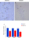
Figure 8.. Intracellular Aβ in adjuvant-treated (ID 174) (A) and immunised (ID 382) (B) lemur brains.
Intracellular Aβ labelling (arrows) in numerous pyramidal neurons of hippocampus of one adjuvant treated animal (A, ID247) and in few pyramidal neurons of hippocampus in one K6Aβ1–30 -treated (B, ID355); C: Intracellular Aβ quantification in frontal, parietal, occipital cortex in adjuvant (n=16) and K6Aβ1–30 - immunised lemurs (n=16). **p < 0.01. Bar: 30 μm.
Figure 9.
Figure 9.. Tau levels in mouse lemur brains.
Immunisation had no effect on total tau (Tau-5) (A,B), soluble phospho-tau PHF-1 (C,D), CP13 (E,F) ) or their ratios (G) on western blots of K6Aβ1–30 mouse lemur brain homogenates compared to adjuvant treated animals. Representative blots are shown on the left. Each bar represents the group average +/− SEM.
Figure 10.
Figure 10.. Tau histology in mouse lemur brain.
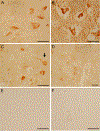
A-B: CP13+ neurons in hippocampus of immunised (A) (ID 355) or adjuvant treated (B)(ID 129) animals with Aβ plaques. Pathological tau in neurons of the piriform cortex of the adjuvant-treated animal (ID 129) evidenced by PHF1 showing tangle (arrow) (C) or AT100 (D) antibodies. E -F: control sections : omission of the primary antibody for the adjuvant treated animal (ID129, E) and for the K6Aβ1–30-treated animal (ID355, F). Bars: 30 μm.
Figure 11.
Figure 11.. Microglial cells evidenced by anti-ferritin antibody in mouse lemur parietal cortex.
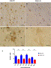
(A-B) Microglial cells stained by ferritin antibody in an adjuvant treated animal (ID129, A) and in an immunised animal (ID355, B). (C-D) Microglial cells in the vicinity of amyloid plaques; double staining Aβ1–42 (in brown) / ferritin (in black) in an adjuvant-treated lemur with amyloid plaques (ID 129, C) and a K6Aβ1–30-immunised lemur with amyloid plaques (ID 355, D). Bar : 30 μm. (E) Ferritin quantification in adjuvant-treated (n=16) and K6Aβ1–30-immunised (n=16) lemurs. Immunised animals had less ferritin staining in frontal (FC), parietal (PC) occipital cortex (OC) and hippocampus (Hpc), compared to adjuvant treated lemurs (**p < 0.01).
Figure 12.
Figure 12.. Astrogliosis in mouse lemur brain with or without Aβ plaques.
GFAP labelling in mouse lemur brain with Aβ plaques (A, E : ID 129), (B, F) (ID 355). E, F : Double staining GFAP (black)/Aβ1–42 (brown) showing astrocytes in the vicinity of Aβ plaques. C-D GFAP labelling in animals without amyloid plaques.C :ID 327, D :ID 385. Bar: 30 μm

References

    1. Alzforum 2022a. : https://www.alzforum.org/therapeutics
    1. Alzforum 2022b. :https://www.alzforum.org/news/research-news/finally-big-win-all-outcomes...).
    1. Asuni AA; Boutajangout A; Scholtzova H; Knudsen E; Li YS; Quartermain D; Frangione B; Wisniewski T; Sigurdsson EM Vaccination of Alzheimer’s Model Mice with Abeta Derivative in Alum Adjuvant Reduces Abeta Burden without Microhemorrhages. Eur J Neurosci 2006, 24 (9), 2530–2542. 10.1111/j.1460-9568.2006.05149.x. - DOI - PMC - PubMed
    1. Bastrup J; Hansen KH; Poulsen TBG, Kastaniegaard K; Asuni AA; Christensen S; Belling D; Helboe L; Stensballe A; Volbracht C. Anti-Aβ Antibody Aducanumab Regulates the Proteome of Senile Plaques and Closely Surrounding Tissue in a Transgenic Mouse Model of Alzheimer’s Disease. J Alzheimers Dis. 2021;79(1):249–265. doi: 10.3233/JAD-200715. PMID: 33252074. - DOI - PubMed
    1. Bons N; Mestre N; Petter A. Senile Plaques and Neurofibrillary Changes in the Brain of an Aged Lemurian Primate, Microcebus Murinus. Neurobiol Aging 1992, 13 (1), 99–105. 10.1016/0197-4580(92)90016-q. - DOI - PubMed

Publication types