. 2023 Jan;11(1):e005878.
doi: 10.1136/jitc-2022-005878.

CD4 CAR-T cells targeting CD19 play a key role in exacerbating cytokine release syndrome, while maintaining long-term responses

Affiliations

CD4 CAR-T cells targeting CD19 play a key role in exacerbating cytokine release syndrome, while maintaining long-term responses

Camilla Bove et al. J Immunother Cancer. 2023 Jan.

Abstract

Background: To date, T cells redirected with CD19-specific chimeric antigen receptors (CAR) have gained impressive success in B-cell malignancies. However, treatment failures are common and the occurrence of severe toxicities, such as cytokine release syndrome (CRS), still limits the full exploitation of this approach. Therefore, the development of cell products with improved therapeutic indexes is highly demanded.

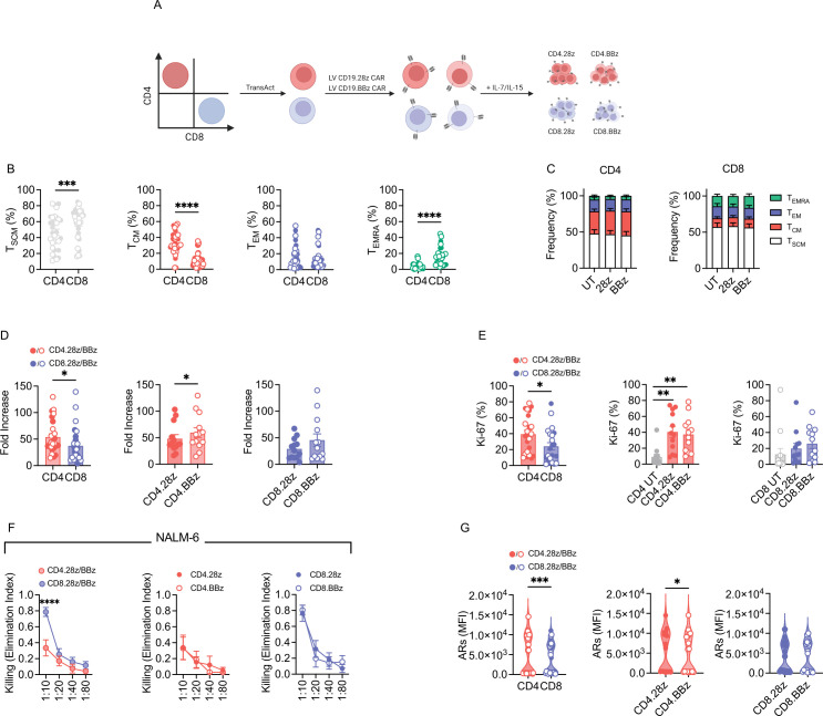
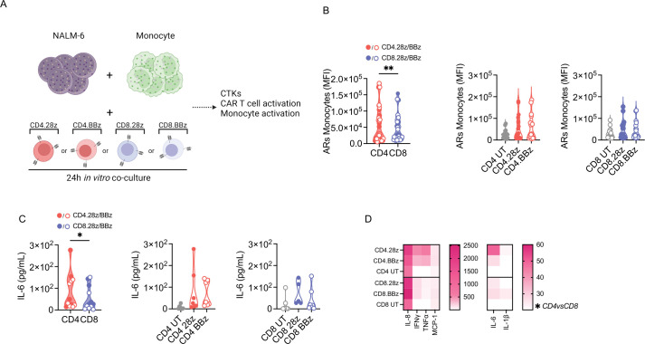
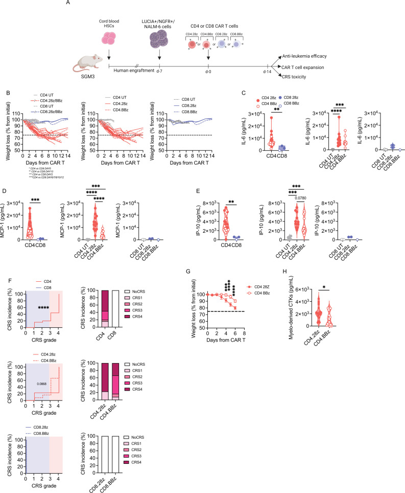
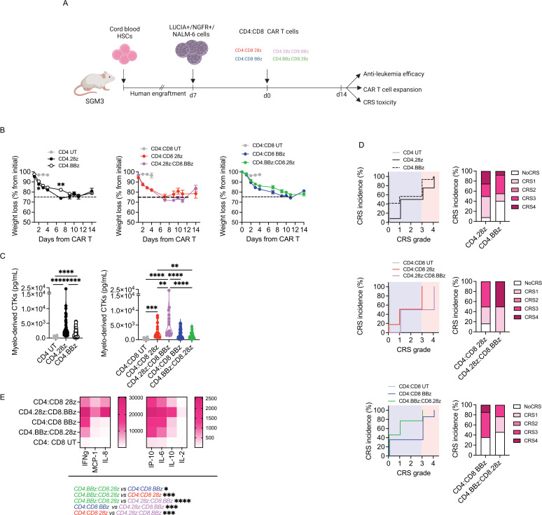
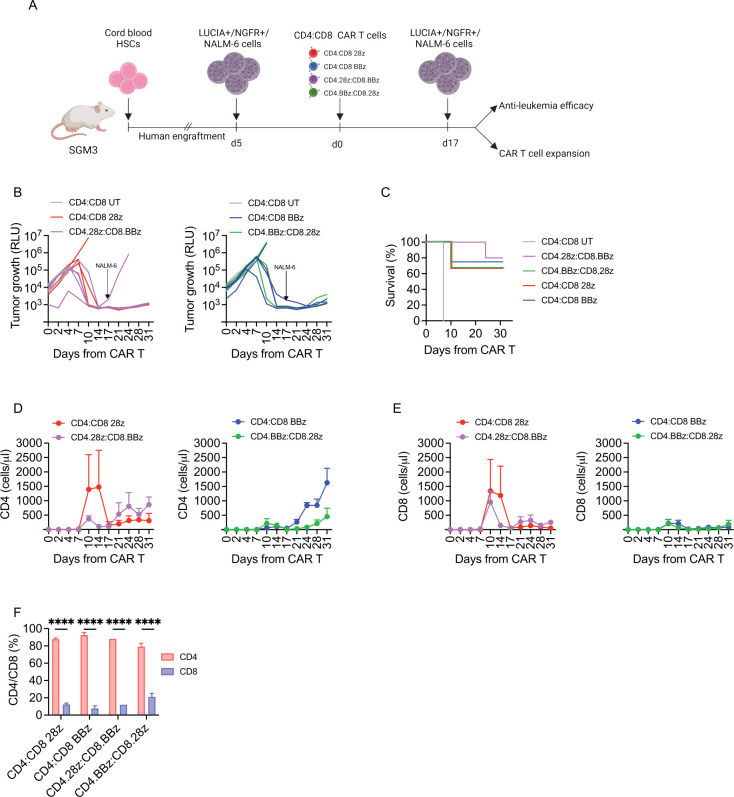
Methods: In this project, we investigated how CD4 and CD8 populations cooperate during CD19 CAR-T cell responses and what is their specific role in CRS development. To this aim, we took advantage of immunodeficient mice reconstituted with a human immune system (HuSGM3) and engrafted with the B-cell acute lymphoblastic leukemia cell line NALM-6, a model that allows to thoroughly study efficacy and toxicity profiles of CD19 CAR-T cell products.

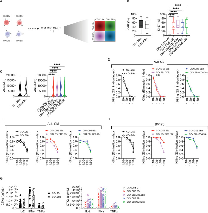
Results: CD4 CAR-T cells showed superior proliferation and activation potential, which translated into stronger stimulation of myeloid cells, the main triggers of adverse events. Accordingly, toxicity assessment in HuSGM3 mice identified CD4 CAR-T cells as key contributors to CRS development, revealing a safer profile when they harbor CARs embedded with 4-1BB, rather than CD28. By comparing differentially co-stimulated CD4:CD8 1:1 CAR-T cell formulations, we observed that CD4 cells shape the overall expansion kinetics of the infused product and are crucial for maintaining long-term responses. Interestingly, the combination of CD4.BBz with CD8.28z CAR-T cells resulted in the lowest toxicity, without impacting antitumor efficacy.

Conclusions: Taken together, these data point out that the rational design of improved adoptive T-cell therapies should consider the biological features of CD4 CAR-T cells, which emerged as crucial for maintaining long-term responses but also endowed by a higher toxic potential.

Keywords: CD4-Positive T-Lymphocytes; CD8-Positive T-Lymphocytes; Cytokines; Immunotherapy; Receptors, Chimeric Antigen.


Conflict of interest statement

Competing interests: CBon received research support from Intellia Therapeutics. ABo is currently an employee of AstraZeneca. His contribution to this work relates to the period 2016–2017 when he was an employee of Vita-Salute San Raffaele University. The other authors declare no competing interests.

Figures

Figure 1
Figure 1
CD4 CAR-T cells display greater activation and proliferation potential. (A) Schematic of CD4 and CD8 CAR-T cell manufacturing. CD4 and CD8 subsets were selected through magnetic sorting, activated, transduced with a lentiviral vector encoding CD19.28z or CD19.BBz CARs and expanded with IL-7/IL-15. (B) Memory phenotype at the end of manufacturing (n=16). (C) Memory phenotype at the end of manufacturing based on CAR co-stimulation (n=8). Untransduced (UT) CD4 and CD8 T cells were used as controls. (D) CAR-T cell fold expansion at the end of manufacturing (n=13). (E) CAR-T cell proliferation after 4-day co-culture with CD19+ tumor cells measured by intracellular staining of Ki-67 (n=3 donors against BV173 and ALL-CM, n=6 donors against NALM-6). (F) Killing activity expressed as Elimination Index and measured after 4-day co-culture with tumor cells at different effector/target (E:T) ratios (n=8). (G) CAR-T cell activation measured as mean fluorescence intensity (MFI) of CD69 and CD25 activation receptors (ARs) 1 day after co-culture with tumor cells (n=3). Data are represented as mean±SEM with overlapping scattered values. *p<0.05, **p<0.01, ***p<0.001, ****p<0.0001 by paired t-test or two-way analysis of variance. CAR, chimeric antigen receptors; IL, interleukin; TCM, central memory T cells; TSCM, stem memory; TEM, effector memory; TEMRA, effector subtypes.
Figure 2
Figure 2
CD4 CAR-T cells are more potent in triggering monocyte activation and cytokine release. (A) Schematic of tripartite co-cultures including NALM-6 leukemia cells, CAR-T cells and autologous monocytes. UT CD4 and CD8 T cells were used as controls. CTKs, cytokines. (B) Activation receptors upregulation (ARs: CD54, CD86, HLA-DR) on monocytes expressed as MFI 1 day after plating (n=6). (C) IL-6 production and (D) heatmap visualization of cytokine release 1 day after plating. Data are represented as mean±SEM or mean±SEM with overlapping scattered or scaled values according to a graded-color range depending on relative minimum and maximum levels, when referring to the heatmap. *p<0.05, **p<0.01, by paired t test or two-way analysis of variance. HLA-DR, human leukocyte antigen-DR isotype; IFN, interferon; IL, interleukin; MFI, mean fluorescence intensity; TNF, tumor necrosis factor; UT, untransduced.
Figure 3
Figure 3
CD4 CAR-T cells exacerbate CRS, especially with the CD28 design. (A) SGM3 mice were reconstituted with human hematopoietic stem/precursor cells (HuSGM3) and injected with Lucia+/NGFR+/NALM-6 leukemia cells. After reaching a high tumor burden, mice were treated with high T-cell doses. Only mice that responded to therapy were included in CRS analysis: CD4.28z (n=13), CD4.BBz (n=12), CD8.28z (n=2) and CD8.BBz (n=1). CD4 UT and CD8 UT (n=6) were used as control. (B) Weight loss at different time points. (C) IL-6, MCP-1 (D) and IP-10 (E) serum levels 4 days after treatment. (F) CRS grading. Left panels: Kaplan-Meier curves. Right panels: Histograms summarizing CRS grading. (G) After reaching a high tumor burden, mice were treated with low doses of CD4.28z and CD4.BBz (n=4) and monitored for weight loss at different time points. (H) Myelo-derived cytokines (IL-6, IP-10, MCP-1) serum levels 4 days after treatment. Data are represented as box and violin plots, mean±SEM together with scaled values according to a graded-color range depending on relative minimum and maximum levels, when referring to the heatmap. *p<0.05, **p<0.01, ***p<0.001, ****p<0.0001, by two-way analysis of variance, unpaired t-test and Gehan-Breslow-Wilcoxon test. CAR, chimeric antigen receptors; CRS, cytokine release syndrome; HSC, hematopoietic stem cell; MCP-1, monocyte chemoattractant protein-; IP-10, interferon γ-induced protein 10; IL, interleukin; UT, untransduced.
Figure 4
Figure 4
CD4:CD8 CAR-T cells display similar in vitro activity. (A) Schematic of CD4:CD8 CAR-T cell manufacturing. The conditions previously employed were then formulated at a 1:1 ratio as follows: CD4:CD8 28z, CD4:CD8 BBz, CD4.28z:CD8.BBz and CD4.BBz:CD8.28z. (B) CAR-T cell proliferation after 4-day co-culture with tumor cells, measured by intracellular staining of Ki-67 (n=3 donors against BV173, n=4 against ALL-CM and n=6 against NALM-6). UT CD4 and CD8 T cells were used as controls. (C) CAR-T cell activation measured as MFI of CD69 and CD25 ARs 1 day after co-culture with tumor cells (n=3). Killing activity expressed as Elimination Index and measured by co-culturing CAR-T cells with (D) NALM-6 (n=3), (E) ALL-CM (n=4) and (F) BV173 (n=3) tumor cells for 4 days at different E:T ratios. (G) Cytokine production after 24-hour co-culture of T cells with tumor cells at a 1:10 E:T ratio (n=3 donors against NALM-6 and ALL-CM, n=1 against BV173). Data are represented as mean±SEM with overlapping scattered values. ****p<0.0001 by paired t-test or two-way analysis of variance. ARs, activation receptors; CAR, chimeric antigen receptors; CTKs, cytokines; E:T, effector:target; IFN, interferon; IL, interleukin; MFI, mean fluorescence intensity; TNF, tumor necrosis factor; UT, untransduced.
Figure 5
Figure 5
CD4.BBz:CD8.28z displays slightly reduced CRS incidence and severity. (A) SGM3 mice were reconstituted with human hematopoietic stem/precursor cells (HuSGM3) and injected with Lucia+/NGFR+/NALM-6 leukemia cells. After reaching a high tumor burden, mice were treated with high doses of CD4:CD8 UT (n=8), CD4:CD8 28z (n=6), CD4:CD8 BBz (n=6), CD4.28z:CD8.BBz (n=14), CD4.BBz:CD8.28z (n=13). (B) Weight loss at different time points. (C) Myelo-derived cytokine (IL-6, IP-10, MCP-1) serum levels 4 days after treatment. (D) CRS grading. Left panels: Kaplan-Meier curves. Right panels: Histograms summarizing CRS grading. (E) Heatmap visualization of serum cytokine levels 4 days after CAR-T cells infusion. Data are represented as mean±SEM with overlapping scattered values and box and violin plots. *p<0.05, **p<0.01, ***p<0.001, ****p<0.0001, by two-way analysis of variance, unpaired t-test and Gehan-Breslow-Wilcoxon test. CAR, chimeric antigen receptors; CRS, cytokine release syndrome; CTKs, cytokines; HSC, hematopoietic stem cell; MCP-1, monocyte chemoattractant protein-; IP-10, interferon γ-induced protein 10; IFN, interferon; IL, interleukin; UT, untransduced.
Figure 6
Figure 6
Expansion profile and kinetic are guided by CD4 CAR-T cells. (A) Schematic of HuSGM3 injected with Lucia+/NGFR+/NALM-6 leukemia cells and treated with low doses of CD4:CD8 UT (n=2), CD4:CD8 28z (n=3), CD4:CD8 BBz (n=4), CD4.28z:CD8.BBz (n=5), CD4.BBz:CD8.28z (n=3) after reaching a mid-tumor burden. (B) NALM-6 bioluminescence signal at different time points after treatment. (C) Kaplan-Meier survival analysis. (D) CD4 and (E) CD8 CAR-T cell expansion peak at different time points after treatment. (F) CD4/CD8 frequency in the bone marrow of surviving mice at sacrifice (n=2). Data are represented as mean±SEM with individual and overlapping scattered values. ****p<0.0001, two-way analysis of variance, by unpaired t-test and Mantel-Cox 2-sided log-rank test. CAR, chimeric antigen receptors; HSC, hematopoietic stem cell; RLU, relative bioluminescent units; UT, untransduced.

References

    1. Locke FL, Ghobadi A, Jacobson CA, et al. . Long-term safety and activity of axicabtagene ciloleucel in refractory large B-cell lymphoma (ZUMA-1): a single-arm, multicentre, phase 1-2 trial. Lancet Oncol 2019;20:31–42. 10.1016/S1470-2045(18)30864-7 - DOI - PMC - PubMed
    1. Schuster SJ, Bishop MR, Tam CS, et al. . Tisagenlecleucel in adult relapsed or refractory diffuse large B-cell lymphoma. N Engl J Med 2019;380:45–56. 10.1056/NEJMoa1804980 - DOI - PubMed
    1. Abramson JS, Palomba ML, Gordon LI, et al. . Lisocabtagene maraleucel for patients with relapsed or refractory large B-cell lymphomas (TRANSCEND NHL 001): a multicentre seamless design study. Lancet 2020;396:839–52. 10.1016/S0140-6736(20)31366-0 - DOI - PubMed
    1. Wang M, Munoz J, Goy A, et al. . KTE-X19 CAR T-cell therapy in relapsed or refractory mantle-cell lymphoma. N Engl J Med 2020;382:1331–42. 10.1056/NEJMoa1914347 - DOI - PMC - PubMed
    1. Munshi NC, Anderson LD, Shah N, et al. . Idecabtagene Vicleucel in relapsed and refractory multiple myeloma. N Engl J Med 2021;384:705–16. 10.1056/NEJMoa2024850 - DOI - PubMed

Publication types