Case Reports
. 2022 Dec 5;15(12):e252080.
doi: 10.1136/bcr-2022-252080.

Total knee replacement in a transtibial amputee

Affiliations
Case Reports

Total knee replacement in a transtibial amputee

Ahmed Elsayed et al. BMJ Case Rep. .

Abstract

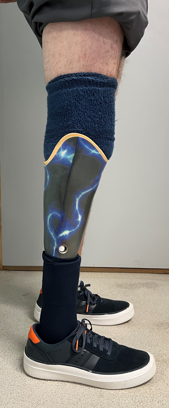
We present the case of a man in his 60s with a transtibial amputation (TTA) undergoing total knee replacement (TKR) for symptomatic osteoarthritis (OA). It is unusual to develop OA in the ipsilateral knee to TTA; and while it is postulated that this is because patients preferentially load their unaffected limb to protect the TTA-sided knee, there is also the ability to offload specific knee compartments through prosthetic adjustment. When planning TKR in such patients, it is important to consider several technical challenges in order to prevent a poor outcome. The literature is sparse with evidence to guide decision-making, and this case report and literature review aims to summarise our preoperative planning and intraoperative technique, which ultimately resulted in a good outcome.

Keywords: Orthopaedic and trauma surgery; Orthopaedics.


Conflict of interest statement

Competing interests: None declared.

Figures

Figure 1
Figure 1
Preoperative lateral view of the right knee. This radiograph demonstrates severe arthritic changes with loss of joint space, subchondral sclerosis, and osteophytes. Changes are present in both the tibiofemoral and patellofemoral joints.
Figure 2
Figure 2
Preoperative anteroposterior view of the right knee. This radiograph further illustrates the lack of joint space in both medial and lateral joint compartments. There is severe subchondral sclerosis particularly along the medial femoral and tibial joint surfaces.
Figure 3
Figure 3
Intraoperative photograph of set-up. This image shows the amputated limb resting on the sterile triangle bolster. The set-up allowed easy flexion and extension of the knee throughout surgery.
Figure 4
Figure 4
Intraoperative photograph with implants in situ. This image shows the Zimmer NexGen knee replacement in situ during the procedure prior to closure. It shows access to the joint via the medial parapatellar approach.
Figure 5
Figure 5
Postoperative lateral view radiograph of the right knee. This is a weight-bearing lateral radiograph showing the Zimmer NexGen knee replacement in situ. It shows the articulation of the patella within the intercondylar groove as well as well-fitting femoral and tibial components.
Figure 6
Figure 6
Postoperative anteroposterior view radiograph of the right knee. This is a weight-bearing anteroposterior radiograph showing the Zimmer NexGen knee replacement in the coronal plane.
Figure 7
Figure 7
Alignment anteroposterior full-length radiograph. This is an important view in assessing the entire limb alignment with the prosthesis. It shows good alignment of the knee replacement with the mechanical axis from the hip centre to ankle centre running just medial to the centre of the knee joint.
Figure 8
Figure 8
Anterior view of the patient’s wound and stump at 6 months postoperatively. This is an anterior view of the patient’s knee taken in clinic 6 months following the procedure. The wound is well healed and there are no concerns regarding the stump.
Figure 9
Figure 9
Lateral view of the patient’s wound and stump at 6 months postoperatively. This is a lateral view of the patient’s knee taken in clinic 6 months following the procedure showing the knee at full extension.
Figure 10
Figure 10
Anterior view of the patient standing with prosthesis. This image demonstrates the alignment of the lower limb with the prosthesis in situ at 6 months postoperatively.
Figure 11
Figure 11
Lateral view of the patient standing with prosthesis. This image demonstrates the lateral alignment of the lower limb while weight-bearing with the prosthesis in situ at 6 months postoperatively.

References

    1. Struyf PA, van Heugten CM, Hitters MW, et al. . The prevalence of osteoarthritis of the intact hip and knee among traumatic leg amputees. Arch Phys Med Rehabil 2009;90:440–6. 10.1016/j.apmr.2008.08.220 - DOI - PubMed
    1. Norvell DC, Czerniecki JM, Reiber GE, et al. . The prevalence of knee pain and symptomatic knee osteoarthritis among veteran traumatic amputees and nonamputees. Arch Phys Med Rehabil 2005;86:487–93. 10.1016/j.apmr.2004.04.034 - DOI - PMC - PubMed
    1. Karam MD, Willey M, Shurr DG. Total knee replacement in patients with below-knee amputation. Iowa Orthop J 2010;30:150–2. - PMC - PubMed
    1. Putnis SE, Neri T, Coolican MRJ. Total knee arthroplasty in a transtibial amputee. J Orthop Case Rep 2020;10:15–18. 10.13107/jocr.2020.v10.i03.1728 - DOI - PMC - PubMed
    1. Crawford JR, Coleman N. Total knee arthroplasty in a below-knee amputee. J Arthroplasty 2003;18:662–5. 10.1016/S0883-5403(03)00209-2 - DOI - PubMed

Publication types