

Interactive spinal computed tomography angiography-guided spinal digital subtraction angiography and embolization for thoracolumbar epidural arteriovenous fistulas: illustrative case
- PMID: 36593676
- PMCID: PMC9514287
- DOI: 10.3171/CASE22275
Interactive spinal computed tomography angiography-guided spinal digital subtraction angiography and embolization for thoracolumbar epidural arteriovenous fistulas: illustrative case
Abstract
Background: Spinal digital subtraction angiography (sDSA) is the gold standard for examining spinal arteriovenous fistulas; however, thorough sDSA evaluations of spinal arteriovenous fistulas require a long procedure, which may increase the radiation exposure time.
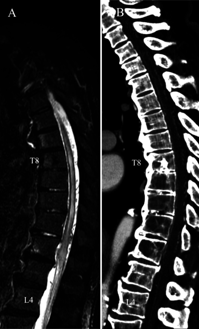
Observations: A 72-year-old man presented with progressive myelopathy due to a spinal epidural arteriovenous fistula. Spinal computed tomography angiography (sCTA) showed an epidural arteriovenous fistula fed by the left L3 segmental artery. To prepare for sDSA, the sCTA images were modified to mark the segmental artery bifurcations from T5 to L5 with multicolored markers. These modified sCTA images were loaded onto the multiwindow DSA display. The sCTA images were interactively modulated during sDSA. This sCTA-guided sDSA identified 18 segmental arteries within 47 minutes. The total radiation exposure was 1,292 mGy. Subsequently, transarterial embolization resolved the epidural arteriovenous fistula with clinical improvement.
Lessons: Three-dimensional sCTA can provide detailed anatomical information before sDSA. Modified sCTA images with segmental artery bifurcation marking can provide interactive guidance on multipanel DSA displays. sCTA-guided sDSA is useful for accurate catheterization and reduction of procedure time.
Keywords: computed tomography angiography; digital subtraction angiography; spinal epidural arteriovenous fistula.
Conflict of interest statement
Figures


Similar articles
-
Spinal intraosseous arteriovenous fistulas with perimedullary drainage associated with vertebral compression fracture: illustrative case.J Neurosurg Case Lessons. 2022 Jul 25;4(4):CASE22184. doi: 10.3171/CASE22184. eCollection 2022 Jul 25. J Neurosurg Case Lessons. 2022. PMID: 36046270 Free PMC article.
-
Spinal intraarterial computed tomography angiography as an effective adjunct for spinal angiography.J Neurosurg Spine. 2015 Sep;23(3):360-7. doi: 10.3171/2014.12.SPINE14584. Epub 2015 May 29. J Neurosurg Spine. 2015. PMID: 26023900
-
Cervical spinal epidural arteriovenous fistula with coexisting spinal anterior spinal artery aneurysm presenting as subarachnoid hemorrhage--case report.J Stroke Cerebrovasc Dis. 2014 Nov-Dec;23(10):e461-e465. doi: 10.1016/j.jstrokecerebrovasdis.2014.07.012. Epub 2014 Oct 3. J Stroke Cerebrovasc Dis. 2014. PMID: 25284720
-
Spinal ventral epidural arteriovenous fistulas of the lumbar spine: angioarchitecture and endovascular treatment.Neuroradiology. 2013 Feb;55(3):327-36. doi: 10.1007/s00234-012-1130-9. Epub 2013 Jan 11. Neuroradiology. 2013. PMID: 23306215 Free PMC article. Review.
-
Dural arteriovenous fistula of the lateral foramen magnum region: A review.Interv Neuroradiol. 2018 Aug;24(4):425-434. doi: 10.1177/1591019918770768. Epub 2018 May 4. Interv Neuroradiol. 2018. PMID: 29726736 Free PMC article. Review.
Cited by
-
Robotic spinal angiography: A single-center experience.Interv Neuroradiol. 2024 Aug 8:15910199241272515. doi: 10.1177/15910199241272515. Online ahead of print. Interv Neuroradiol. 2024. PMID: 39113603 Free PMC article.
References
-
- Yamaguchi S, Takemoto K, Takeda M, et al. The position and role of four-dimensional computed tomography angiography in the diagnosis and treatment of spinal arteriovenous fistulas. World Neurosurg. 2017;103:611–619. - PubMed
-
- Zhou G, Li MH, Lu C, et al. Dynamic contrast-enhanced magnetic resonance angiography for the localization of spinal dural arteriovenous fistulas at 3T. J Neuroradiol. 2017;44(1):17–23. - PubMed
LinkOut - more resources
Full Text Sources
