

Screening of COVID-19 Based on GLCM Features from CT Images Using Machine Learning Classifiers
- PMID: 36593973
- PMCID: PMC9798374
- DOI: 10.1007/s42979-022-01583-2
Screening of COVID-19 Based on GLCM Features from CT Images Using Machine Learning Classifiers
Abstract
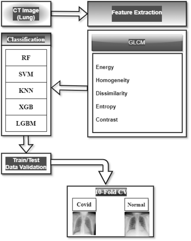
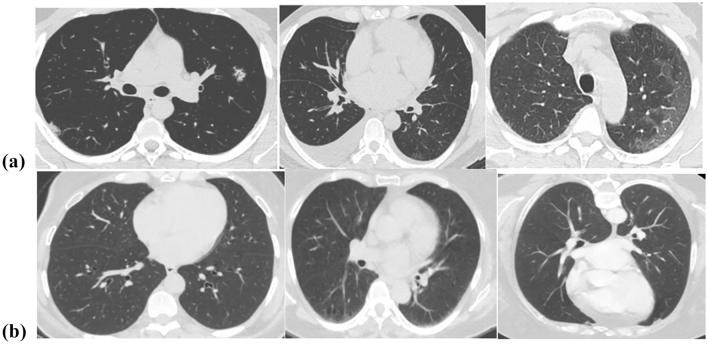
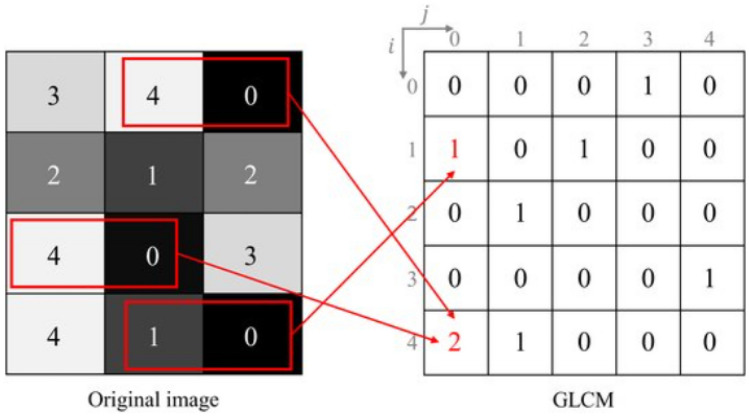
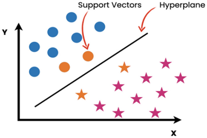
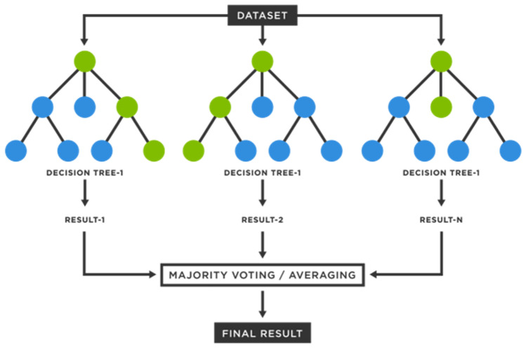
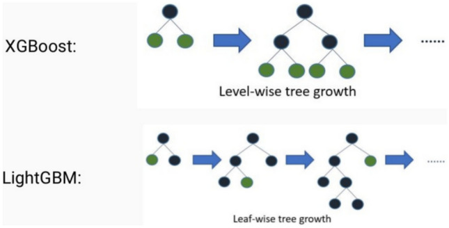
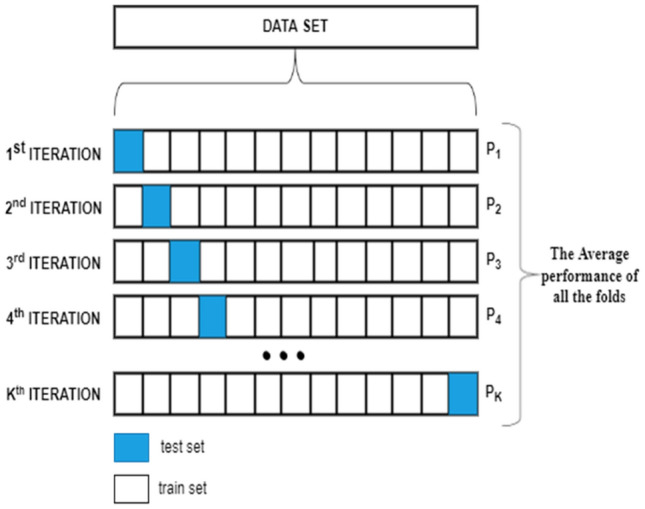
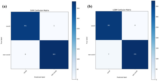
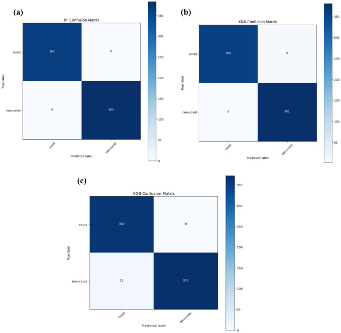
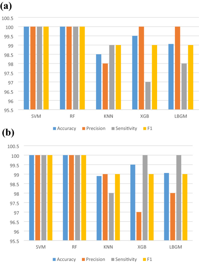
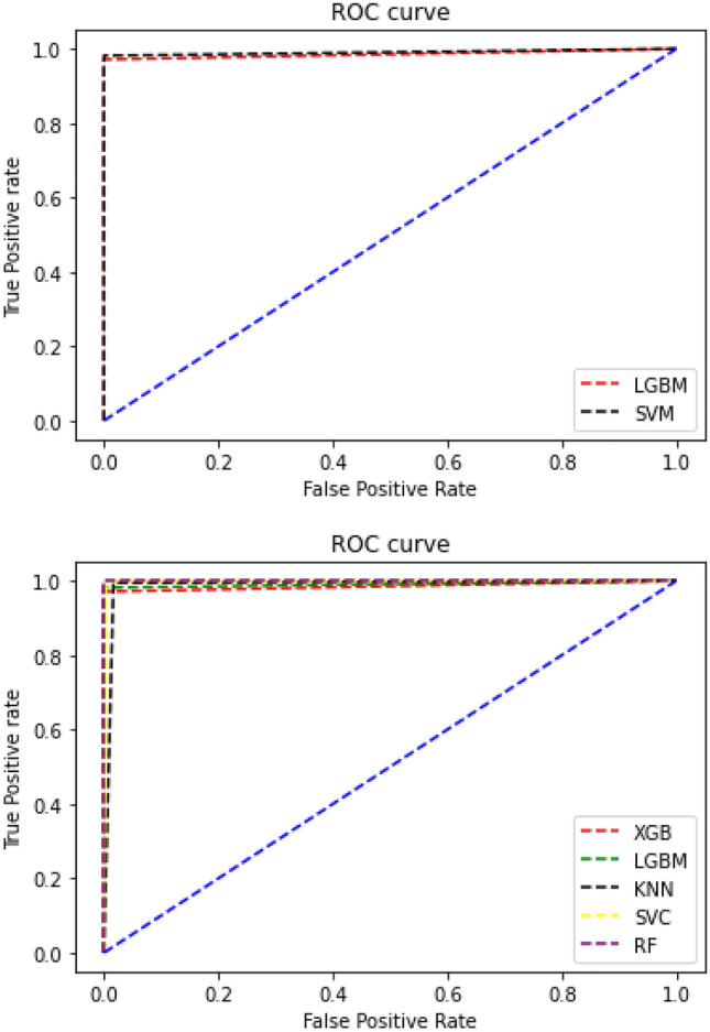
In healthcare, the decision-making process is crucial, including COVID-19 prevention methods should include fast diagnostic methods. Computed tomography (CT) is used to diagnose COVID patients' conditions. There is inherent variation in the texture of a CT image of COVID, much like the texture of a CT image of pneumonia. The process of diagnosing COVID images manually is difficult and challenging. Using low-resolution images and a small COVID dataset, the extraction of discriminant characteristics and fine-tuning of hyperparameters in classifiers provide challenges for computer-assisted diagnosis. In radiomics, quantitative image analysis is frequently used to evaluate the prognosis and diagnose diseases. This research tests an ML model built on GLCM features collected from chest CT images to screen for COVID-19. In this study, Support Vector Machines, K-nearest neighbors, Random Forest, and XGBoost classifiers are used together with LBGM. Tuning tests were used to regulate the hyperparameters of the model. With cross-validation, tenfold results were obtained. Random Forest and SVM were the best classification methods for GLCM features with an overall accuracy of 99.94%. The network's performance was assessed in terms of sensitivity, accuracy, and specificity.
Keywords: COVID-19; Feature extraction; GLCM; LGBM; Machine learning; SVM.
© The Author(s), under exclusive licence to Springer Nature Singapore Pte Ltd 2022, Springer Nature or its licensor (e.g. a society or other partner) holds exclusive rights to this article under a publishing agreement with the author(s) or other rightsholder(s); author self-archiving of the accepted manuscript version of this article is solely governed by the terms of such publishing agreement and applicable law.
Conflict of interest statement
Conflict of InterestThe authors declare that they have no known competing financial interests or personal relationships that could have appeared to influence the work reported in this paper.
Figures
Similar articles
-
Computer-aided diagnosis of COVID-19 from chest X-ray images using histogram-oriented gradient features and Random Forest classifier.Multimed Tools Appl. 2022;81(28):40451-40468. doi: 10.1007/s11042-022-13183-6. Epub 2022 May 10. Multimed Tools Appl. 2022. PMID: 35572385 Free PMC article.
-
Differentiating novel coronavirus pneumonia from general pneumonia based on machine learning.Biomed Eng Online. 2020 Aug 19;19(1):66. doi: 10.1186/s12938-020-00809-9. Biomed Eng Online. 2020. PMID: 32814568 Free PMC article.
-
Differentiation of fat-poor angiomyolipoma from clear cell renal cell carcinoma in contrast-enhanced MDCT images using quantitative feature classification.Med Phys. 2017 Jul;44(7):3604-3614. doi: 10.1002/mp.12258. Epub 2017 Jun 9. Med Phys. 2017. PMID: 28376281
-
Biphasic majority voting-based comparative COVID-19 diagnosis using chest X-ray images.Expert Syst Appl. 2023 Apr 15;216:119430. doi: 10.1016/j.eswa.2022.119430. Epub 2022 Dec 21. Expert Syst Appl. 2023. PMID: 36570382 Free PMC article. Review.
-
Machine learning techniques for CT imaging diagnosis of novel coronavirus pneumonia: a review.Neural Comput Appl. 2022 Sep 19:1-19. doi: 10.1007/s00521-022-07709-0. Online ahead of print. Neural Comput Appl. 2022. PMID: 36159188 Free PMC article. Review.
References
-
- Al-Karawi D, Al-Zaidi S, Polus N, Jassim S. Machine learning analysis of chest CT scan images as a complementary digital test of coronavirus (COVID-19) patients. MedRxiv. 2020.
LinkOut - more resources
Full Text Sources