. 2023 Jan 1;19(1):258-280.
doi: 10.7150/ijbs.66839. eCollection 2023.

Validation of ESM1 Related to Ovarian Cancer and the Biological Function and Prognostic Significance

Affiliations

Validation of ESM1 Related to Ovarian Cancer and the Biological Function and Prognostic Significance

Yu-Kun Li et al. Int J Biol Sci. .

Abstract

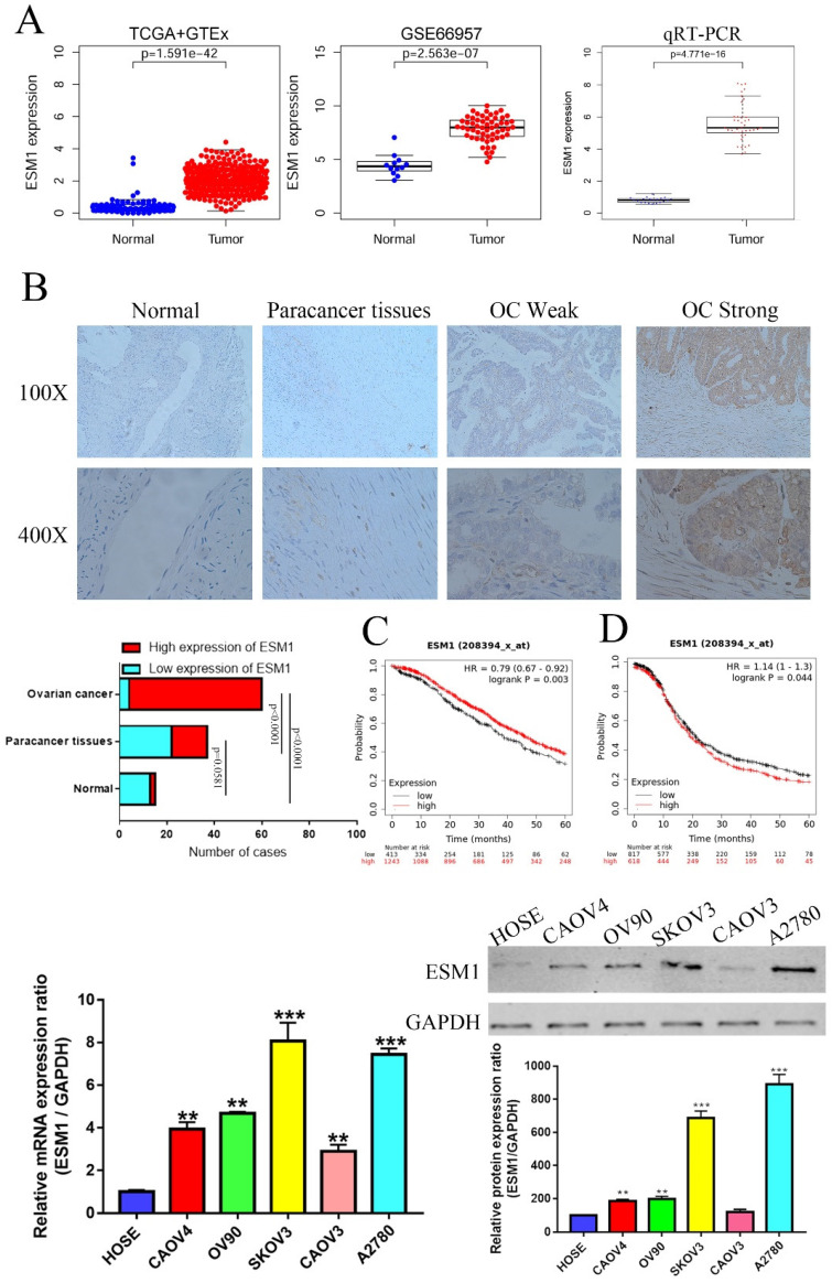
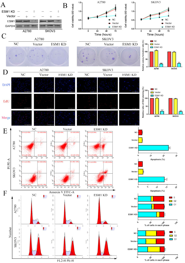
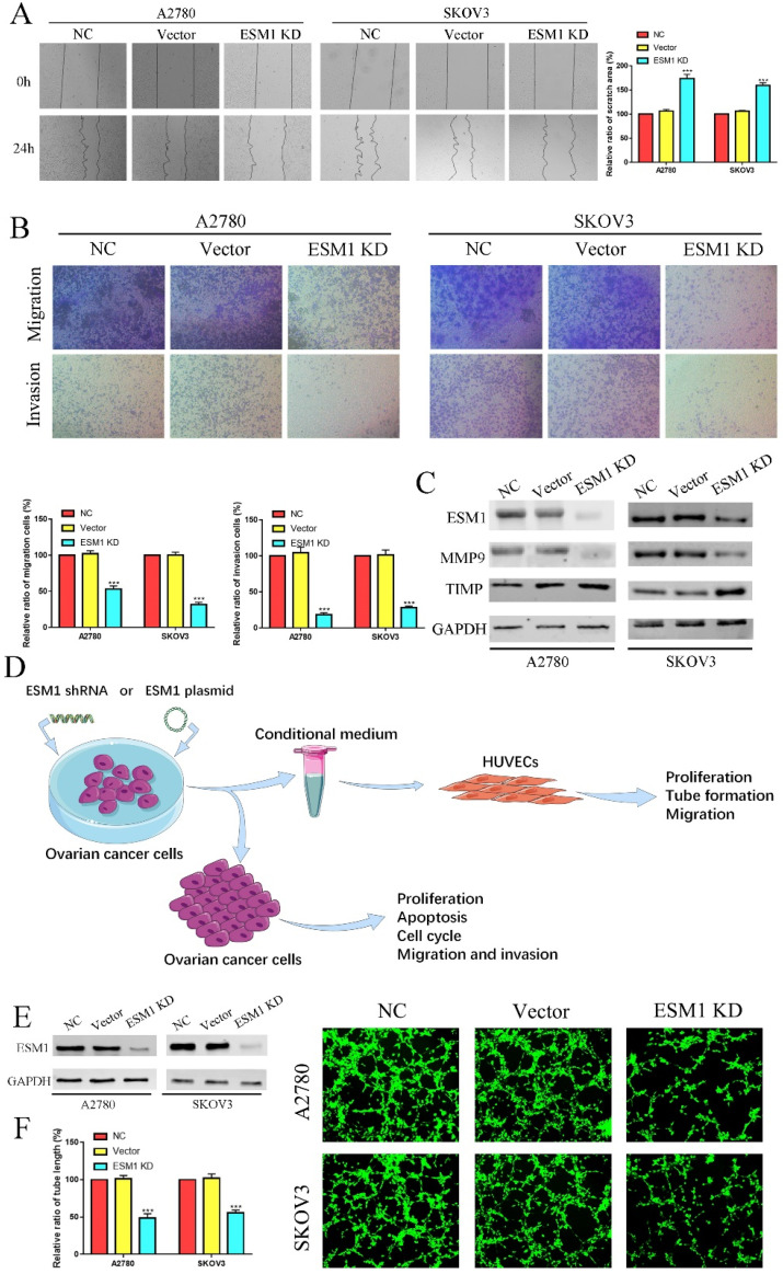
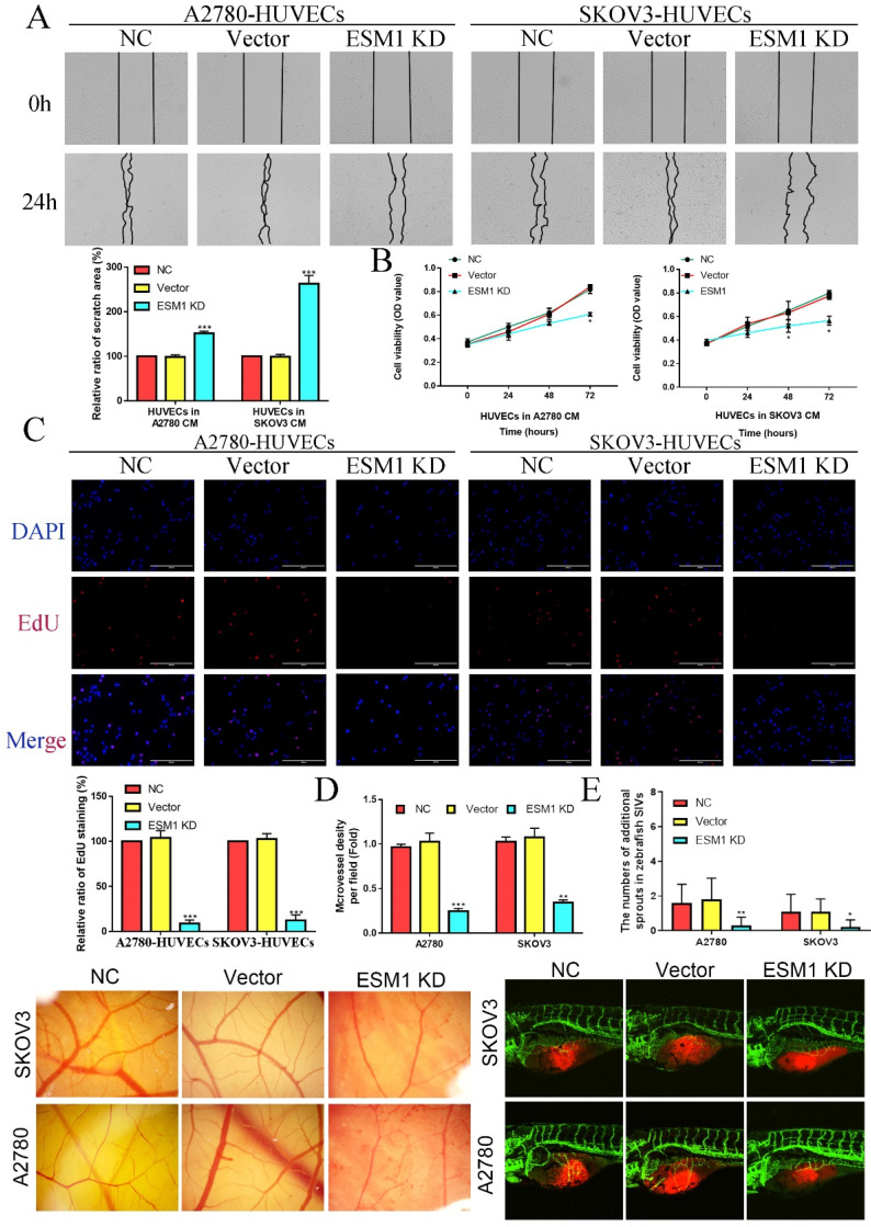
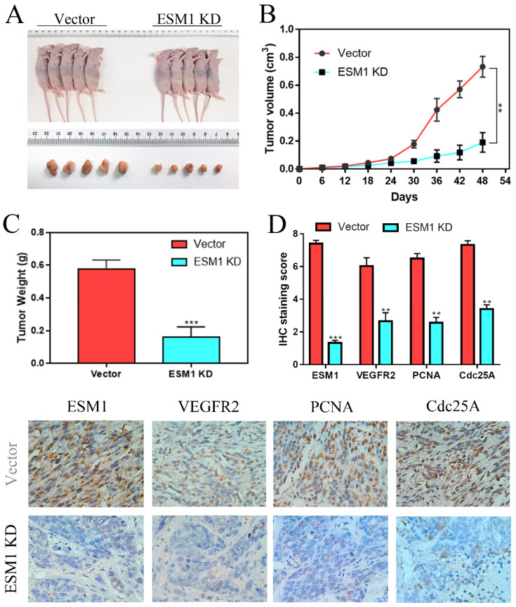
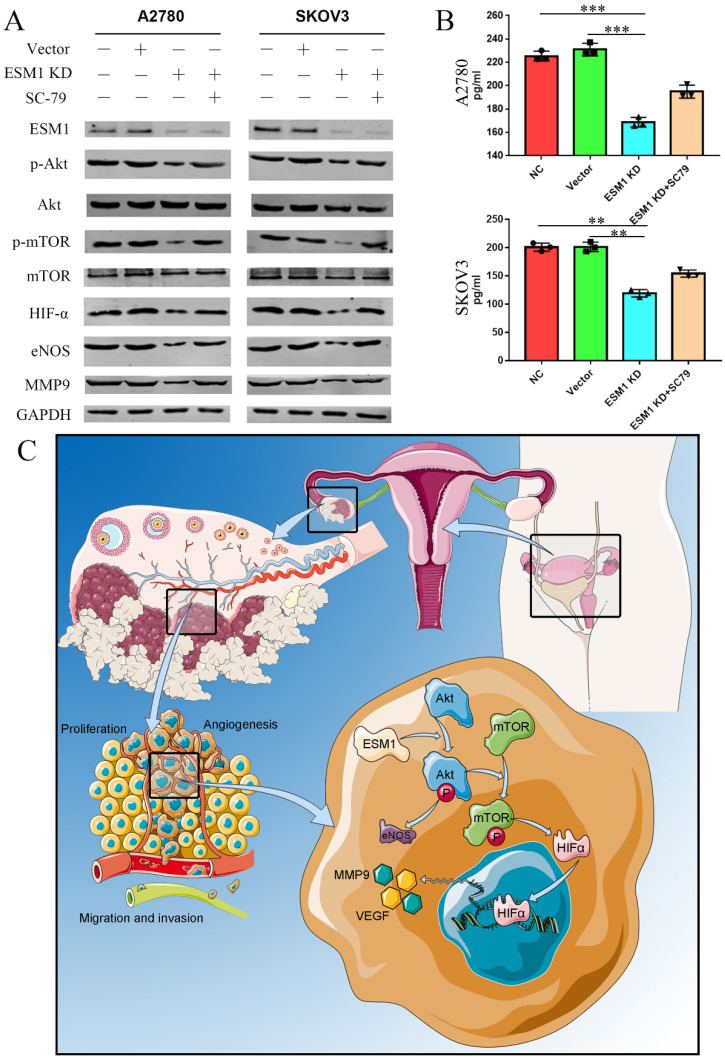
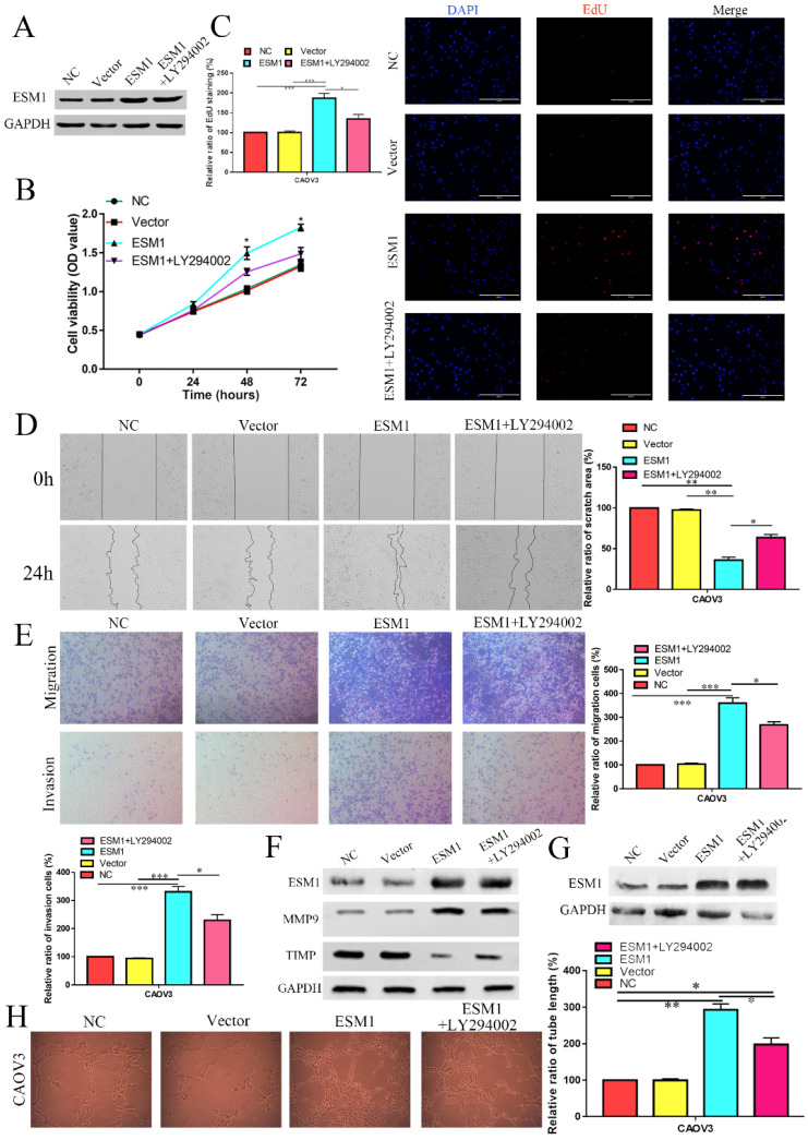
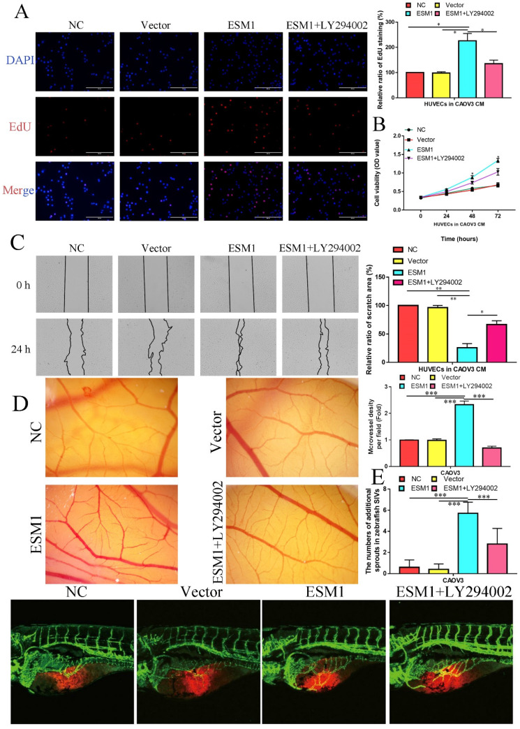
Background: Ovarian cancer (OC), a serious gynecological malignant disease, remains an enormous challenge in early diagnosis and medical treatment. Based on the GEO and TCGA databases in R language, endothelial cell-specific molecule 1 (ESM1) was confirmed separately with the bioinformatic analysis tool. ESM1 has been demonstrated to be upregulated in multiple cancer types, but the oncogenic mechanism by which ESM1 promotes OC is still largely unknown. Methods: In this study, we used WGCNA and random survival forest variable screening to filter out ESM1 in OC differentially expressed genes (DEGs). Next, we confirmed the mRNA and protein levels of ESM1 in OC samples via PCR and IHC. The correlation between the ESM1 level and clinical data of OC patients was further confirmed, including FIGO stage, lymph node metastasis, and recurrence. The role of ESM1 in OC development was explored by several functional experiments in vivo and in vitro. Then, the molecular mechanisms of ESM1 were further elucidated by bioinformatic end experimental analysis. Results: ESM1 was significantly upregulated in OC and was positively correlated with PFS but negatively correlated with OS. ESM1 knockdown inhibited cell proliferation, apoptosis escape, the cell cycle, angiogenesis, migration and invasion in multiple experiments. Moreover, GSVA found that ESM1 was associated with the Akt pathway, and our results supported this prediction. Conclusion: ESM1 was closely correlated with OC development and progression, and it could be considered a novel biomarker and therapeutic target for OC patients.

Keywords: Akt/mTOR pathway; Bioinformatic analysis; ESM1; Ovarian cancer; Prognostic marker.


Conflict of interest statement

Competing Interests: The authors have declared that no competing interest exists.

Figures

Figure 1
Figure 1
Program flowchart for this study.
Figure 2
Figure 2
Validation of the hub module via weighted gene coexpression network analysis (WGCNA). DEGs are shown on the heatmap (A) and the volcano plot (B) for the GSE66957 and GSE54388 datasets. The scale-free fit index and the average connectivity of soft threshold power and hierarchical clustering tree of genes based on topological overlap are confirmed for GSE66957 (C) and GSE54388 (D). The correlation of these modules between the normal group and tumor group for GSE66957 (E) and GSE54388 (F). A scatter plot of the turquoise module for GSE669857 (G) and the blue module for GSE54388 (H).
Figure 3
Figure 3
Validation of key genes in OC. A. Correlation between the error rate and number of trees. B. Variable relative importance of the sequencing of three genes. C. GSVA of three key genes, ESM1, CENPH and HIST1H2AE. D. The networks among ESM1, CENPH and HIST1H2AE based on the GeneMANIA database.
Figure 4
Figure 4
Epigenetic modification and genetic alteration of 3 key genes. A. The DNA alteration of 3 key genes. B. Tumor mutation burden of 3 key genes. C. The estimated IC50 values of cisplatin, docetaxel, erlotinib, gefitinib and paclitaxel for 3 key genes. D. The DNA promoter methylation levels of 3 key genes based on the TCGA database.
Figure 5
Figure 5
The expression level and prognostic value of ESM1 in OC patients. A. Identification of the mRNA level of ESM1 based on the TCGA database OC dataset, GEO database GSE66957 dataset and qRT-PCR in OC patients. B. The protein expression level of ESM1 in normal ovary tissue samples, paracancerous OC tissue samples and OC tissue samples. C. The OS analysis of ESM1 based on the KM-plot database. D. PFS analysis of ESM1 based on the KM plot database. E. The mRNA level of ESM1 in multiple OC cell lines and immortalized ovarian epithelial cells. F. ESM1 protein expression in multiple OC cell lines and immortalized ovarian epithelial cells. **P < 0.01, ***P < 0.001 indicates statistical significance compared with the control.
Figure 6
Figure 6
The effect of ESM1 on OC proliferation, cell cycle progression and apoptosis. A. The expression of ESM1 in the NC, vector and ESM1 KD groups of A2780 and SKOV3 cells. The effect of ESM1 KD on A2780 and SKOV3 cells via MTT analysis (B), plate clone formation assay (C), and EdU assay (D). E. The function of ESM1 KD in OC cell apoptosis escape. F. The effect of ESM1 KD on the cell cycle in A2780 and SKOV3 cells. *P < 0.05, **P < 0.01, ***P < 0.001 indicates statistical significance compared with the control.
Figure 7
Figure 7
ESM1 KD impedes OC migration, invasion and angiogenesis. The migration and invasion ability of the NC, vector, and ESM1 knockdown were measured and compared in A2780 and SKOV3 cells by wound healing (A) and Transwell assays (B). C. The level of MMP9 and TIMP of ESM1 KD by shRNA compared with the normal control and vector group by Western blotting in A2780 and SKOV3 cell lines. D. A workflow of cell transfection, conditional medium collection and analyses for tube formation and other experiments. E. The expression of ESM1 in the NC, vector and ESM1 KD groups of A2780 and SKOV3 cells. F. Cell angiogenesis assays were performed in A2780 and SKOV3 cells transfected with the vector or ESM1 shRNA. ***P < 0.001 indicates statistical significance compared with the control.
Figure 8
Figure 8
ESM1 knockdown can repress OC cell angiogenesis in vitro and vivo. A. A2780/SKOV3-conditioned medium from cells transfected with Vector or ESM1 shRNA enhanced the HUVECs migration ability by wound healing analysis. MTT analysis (B) and EdU assay (C) for the effects of OC cell conditioned medium with ESM1 KD on HUVECs proliferation. D. Chicken embryos were incubated with A2780/SKOV3-conditioned medium after ESM1 KD or not. E. The effects of ESM1 shRNA transfection or empty vector plasmid transfection on A2780/SKOV3 angiogenesis in zebrafish model. *P < 0.05, **P < 0.01, ***P < 0.001 indicates statistical significance compared with the control.
Figure 9
Figure 9
ESM1 inhibition can repress OC cell growth in vivo. A. The effects of ESM1 shRNA lentivirus plasmid transfection or empty vector lentivirus plasmid transfection on A2780 growth in vivo. B. The tumor volume and C. tumor weight of these xenografts. D. The expression of ESM1, VEGFA, PCNA and Cdc25A in these xenografts via IHC staining. **P < 0.01, ***P < 0.001 indicates statistical significance compared with the control.
Figure 10
Figure 10
ESM1 accelerates OC development and progression via the Akt/mTOR pathway. A. The expression of ESM1, Akt, p-Akt, mTOR, p-mTOR, HIF-α, eNOS, and MMP9 was confirmed in the NC, vector, ESM1 KD and ESM1 KD with SC-79 (Akt pathway activator) groups by Western blotting. B. The level of VEGF was detected in the NC, vector, ESM1 KD and ESM1 KD with SC-79 (Akt pathway activator) groups by ELISA analysis. C. The potential mechanisms of ESM1 in OC. **P < 0.01, ***P < 0.001 indicates statistical significance compared with the control.
Figure 11
Figure 11
The effect of ESM1 overexpression on the OC biological phenotype via the Akt pathway. A. The expression of ESM1 in the NC, vector, ESM1, and ESM1+LY294002 groups in CAOV3 cells by Western blotting. The effect of ESM1 and ESM1+LY294002 on CAOV3 cell proliferation ability via MTT analysis (B) and EdU assay (C). The effect of ESM1 and ESM1+LY294002 on CAOV3 cell migration and invasion ability via wound healing (D) and Transwell assays (E). F. The levels of MMP9 and TIMP in ESM1 and ESM1+LY294002 cells compared with the normal control and vector groups by Western blotting in CAOV3 cell lines. G. The expression of ESM1 in the NC, vector, ESM1 and ESM1+LY294002 groups in CAOV3 cell lines. H. Angiogenesis assays were performed in CAOV3 cells transfected with vector, ESM1, or ESM1+LY294002. *P < 0.05, **P < 0.01, ***P < 0.001 indicates statistical significance compared with the control.
Figure 12
Figure 12
ESM1 can promote the OC angiogenesis via the Akt pathway in vitro and vivo. The effect of ESM1 or ESM1+LY294002 CAOV3 cell-conditioned medium on HUVECs proliferation ability via EdU analysis (A) and MTT assay (B). C. The effect of ESM1 or ESM1+LY294002 CAOV3 cell-conditioned medium on HUVECs migration ability via wound healing. D. Chicken embryos were incubated with CAOV3-conditioned medium after ESM1 or ESM1+LY294002. E. The effects of ESM1 or ESM1+LY294002 CAOV3 cell on angiogenesis in zebrafish model. *P < 0.05, **P < 0.01, ***P < 0.001 indicates statistical significance compared with the control.

Similar articles

Cited by

References

    1. Siegel RL, Miller KD, Jemal A. Cancer statistics, 2018. CA: A Cancer Journal for Clinicians. 2018;68:7–30. - PubMed
    1. Lee JM, Minasian L, Kohn EC. New strategies in ovarian cancer treatment. Cancer. 2019;125(Suppl 24):4623–9. - PMC - PubMed
    1. Asante DB, Calapre L, Ziman M, Meniawy TM, Gray ES. Liquid biopsy in ovarian cancer using circulating tumor DNA and cells: Ready for prime time? Cancer Lett. 2020;468:59–71. - PubMed
    1. Fan CA, Reader J, Roque DM. Review of Immune Therapies Targeting Ovarian Cancer. Curr Treat Options Oncol. 2018;19:74. - PubMed
    1. Cortez AJ, Tudrej P, Kujawa KA, Lisowska KM. Advances in ovarian cancer therapy. Cancer Chemother Pharmacol. 2018;81:17–38. - PMC - PubMed