. 2023 Jan 1;19(1):156-166.
doi: 10.7150/ijbs.76148. eCollection 2023.

A stapled chromogranin A-derived peptide homes in on tumors that express αvβ6 or αvβ8 integrins

Affiliations

A stapled chromogranin A-derived peptide homes in on tumors that express αvβ6 or αvβ8 integrins

Matteo Monieri et al. Int J Biol Sci. .

Abstract

Rationale: The αvβ6- and αvβ8-integrins, two cell-adhesion receptors upregulated in many tumors and involved in the activation of the latency associated peptide (LAP)/TGFβ complex, represent potential targets for tumor imaging and therapy. We investigated the tumor-homing properties of a chromogranin A-derived peptide containing an RGDL motif followed by a chemically stapled alpha-helix (called "5a"), which selectively recognizes the LAP/TGFβ complex-binding site of αvβ6 and αvβ8. Methods: Peptide 5a was labeled with IRDye 800CW (a near-infrared fluorescent dye) or with 18F-NOTA (a label for positron emission tomography (PET)); the integrin-binding properties of free peptide and conjugates were then investigated using purified αvβ6/αvβ8 integrins and various αvβ6/αvβ8 single - or double-positive cancer cells; tumor-homing, biodistribution and imaging properties of the conjugates were investigated in subcutaneous and orthotopic αvβ6-positive carcinomas of the pancreas, and in mice bearing subcutaneous αvβ8-positive prostate tumors. Results: In vitro studies showed that 5a can bind both integrins with high affinity and inhibits cell-mediated TGFβ activation. The 5a-IRDye and 5a-NOTA conjugates could bind purified αvβ6/αvβ8 integrins with no loss of affinity compared to free peptide, and selectively recognized various αvβ6/αvβ8 single- or double-positive cancer cells, including cells from pancreatic carcinoma, melanoma, oral mucosa, bladder and prostate cancer. In vivo static and dynamic optical near-infrared and PET/CT imaging and biodistribution studies, performed in mice with subcutaneous and orthotopic αvβ6-positive carcinomas of the pancreas, showed high target-specific uptake of fluorescence- and radio-labeled peptide by tumors and low non-specific uptake in other organs and tissues, except for excretory organs. Significant target-specific uptake of fluorescence-labeled peptide was also observed in mice bearing αvβ8-positive prostate tumors. Conclusions: The results indicate that 5a can home to αvβ6- and/or αvβ8-positive tumors, suggesting that this peptide can be exploited as a ligand for delivering imaging or anticancer agents to αvβ6/αvβ8 single- or double-positive tumors, or as a tumor-homing inhibitor of these TGFβ activators.

Keywords: RGD motif; TGFβ; cancer.; chromogranin A; αvβ6 and αvβ8 integrins.


Conflict of interest statement

Competing Interests: AC, AG, and FC are inventors of a patent regarding CgA-derived peptides.

Figures
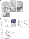
Figure 1
Figure 1
Peptide 5a prevents the adhesion of TRAMP-C2 cells (αvβ8-positive) to plates coated with an anti-αvβ8 antibody or with a peptide 5a-albumin conjugate and inhibits cell-mediated TGFβ activation. (A) Effect of 5a and 2a on the adhesion of TRAMPC-2 cells to PVC-microplates coated with anti-αvβ8 mAb ADWA-11 or mAb 5A8 (a negative control). Cells were mixed with or without the peptides (100 µM) and analyzed by bright-field microscopy after 2h. Representative photographs (10x magnification) are shown. (B) Adhesion of TRAMP-C2 cells to PVC-microplates coated with a 5a-human serum albumin conjugate (5a-HSA), or with a control conjugate made without peptide (*HSA), after 2 h of incubation. Non-adherent cells were removed; adherent cells were stained with crystal violet and quantified by spectrophotometric analysis (A570nm) (mean±SE, n=3). Representative photomicrographs of wells coated with 5a-HSA or *HSA (16.6 µg/ml) are shown. (C) Effect of 5a and 2a on the adhesion of TRAMPC-2 cells to PVC-microplates coated with 5a-HSA (10 µg/ml). Adherent cells were stained with crystal violet and analyzed spectrophotometrically (mean±SE, n=2-3). (D) Effect of 5a and 2a on TGFβ activation by TRAMP-C2 cells. Cells were seeded on cell culture microplates, left to adhere for 3 h and treated with 5a or 2a for 16 h. The amount of active TGFβ in the supernatant was quantified using a bioassay based on HEK-Blue™ TGFβ cells. Cumulative results of two independent experiments are shown (mean±SE, n=4-6 wells). *, P<0.05; **, P<0.01; ***P <0.001 by two-tail t-test.
Figure 2
Figure 2
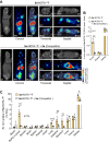
The uptake of 5a-IRDye by subcutaneous BxPC-3 tumor depends on the 5a moiety. (A) Nude mice bearing-BxPC-3 tumors were injected i.v. with the indicated amount of Cys-IRDye, 5a-IRDye, or 5a-IRDye plus an excess of 5a, and imaged at the indicated time points using an IVIS imaging system, 30-35 days after tumor implantation. Pseudocolor images of representative mice for each group of treatment are shown (blue and green arrows indicate tumor and kidney, respectively). Quantification of the fluorescence uptake by BxPC-3 tumors and tumor-to-background ratio are shown in the lower panels (graphs, means ± SE of 3-4 mice/group; *, P<0.05 and **, P<0.01 by ordinary one-way ANOVA of the area under the curve for each tumor using GraphPad Prism software). (B) Similar experiment showing the tumor uptake of 5a-IRDye compared to 5a-Scr-IRDye. Blue and green arrows in pseudocolor images indicate tumor and kidney, respectively, while black arrow indicates the intestine. **, P<0.01; ***P<0.001 by ordinary one-way ANOVA. Black arrow indicates 5a-Scr-IRDye accumulation in the intestine (as confirmed by ex vivo analysis of the isolated organ, not shown).
Figure 3
Figure 3
5a-IRDye homes in on orthotopically implanted BxPC-3 tumors. Nu/nu mice were orthotopically implanted with a Matrigel solution containing BxPC-3 cells or a Matrigel solution without cells (n=3-4 mice). After 18 days, mice were injected i.v. with 5a-IRDye (1.2 nmol), and NIR images were acquired after 24 h. A control mouse, which was not surgically manipulated, was injected with the vehicle and used as a reference for quantification of autofluorescence in the NIR region. (A) Representative fluorescence images of the exposed pancreas of control and tumor-bearing mice injected with or without 5a-IRDye as indicated. (B) Ex vivo fluorescence imaging and photographs of the removed pancreas. Arrow, pancreatic cancer lesions. (C) Ex vivo biodistribution of 5a-IRDye in selected organs. Bars: mean ± SE of 1-4 mice.
Figure 4
Figure 4
Competition of 5a-NOTA-18F uptake by unlabeled peptide 5a in the subcutaneous BxPC-3 tumor model. BxPC-3 tumors-bearing NGS mice were injected i.v. with or without unlabeled 5a (130 nmol/mouse, Competitor) and 10 min later, with 5a-NOTA-18F (~ 3 MBq/animal, i.v.). Radiotracer uptake was assessed after 2 h by whole-body PET/CT (A, B) or with a gamma-counter for biodistribution studies (C). Bars, mean ± SE (n=3 mice). Arrow, BxPC-3 tumor. *, P<0.05, **, P<0.01 and ***P<0.001 by two-tail t-test.

References

    1. Koivisto L, Bi J, Hakkinen L, Larjava H. Integrin alphavbeta6: Structure, function and role in health and disease. Int J Biochem Cell Biol. 2018;99:186–96. - PubMed
    1. Liu H, Wu Y, Wang F, Liu Z. Molecular imaging of integrin alphavbeta6 expression in living subjects. Am J Nucl Med Mol Imaging. 2014;4:333–45. - PMC - PubMed
    1. Elayadi AN, Samli KN, Prudkin L, Liu YH, Bian A, Xie XJ. et al. A peptide selected by biopanning identifies the integrin alphavbeta6 as a prognostic biomarker for nonsmall cell lung cancer. Cancer Res. 2007;67:5889–95. - PubMed
    1. Moore KM, Thomas GJ, Duffy SW, Warwick J, Gabe R, Chou P, Therapeutic targeting of integrin alphavbeta6 in breast cancer. J Natl Cancer Inst. 2014. 106. - PMC - PubMed
    1. Sipos B, Hahn D, Carceller A, Piulats J, Hedderich J, Kalthoff H. et al. Immunohistochemical screening for beta6-integrin subunit expression in adenocarcinomas using a novel monoclonal antibody reveals strong up-regulation in pancreatic ductal adenocarcinomas in vivo and in vitro. Histopathology. 2004;45:226–36. - PubMed

Publication types