

The Correlation Between Morphologic Characteristics of Condyle and Glenoid Fossa with Different Sagittal Patterns of Jaw Assessed by Cone-Beam Computed Tomography
- PMID: 36594548
- PMCID: PMC9885781
- DOI: 10.5152/TurkJOrthod.2022.21136
The Correlation Between Morphologic Characteristics of Condyle and Glenoid Fossa with Different Sagittal Patterns of Jaw Assessed by Cone-Beam Computed Tomography
Abstract
Objective: This study aimed to determine the relationship between the morphologic characteristics of condyle and glenoid fossa in different sagittal skeletal patterns using cone-beam computed tomography.
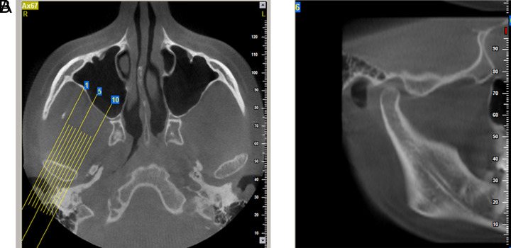
Methods: In this cross-sectional study, the lateral cephalometric and cone-beam computed tomography images of 90 patients were evaluated. The patients were categorized into three equal groups of sagittal skeletal patterns, according to the ANB angle. The greatest anteroposterior and mediolateral diameters of the mandibular condyles, as well as the angle between the long axis of the mandibular condyles and the midsagittal plane, were measured on the axial view of cone-beam computed tomography images. The anterior joint space, superior joint space, posterior joint space, articular eminence inclination, depth of the glenoid fossa, and width of the glenoid fossa were also measured on the central sagittal slices. One-way analysis of variance (ANOVA), Tukey's post hoc test and chi-square test were performed.
Results: Patients with the skeletal Class III had a significantly higher articular eminence inclination, while Class II patients had a lower articular eminence inclination (P = .001). In Class III patients, the depth of the glenoid fossa was greater, and the width of the glenoid fossa was smaller than in the other groups (P < .01). The anterior and posterior joint space did not show any significant differences between the 3 groups.
Conclusion: There were significant differences in some morphological characteristics of the condyle and glenoid fossa in patients with different sagittal skeletal patterns; therefore, this relationship should be considered in the treatment of these patients.
Figures


References
-
- dos Santos PFd. Correlation between sagittal dental classes and sagittal condylar inclination. J Stomat Occ Med. 2013;6(3):96 100. 10.1007/s12548-013-0086-7) - DOI
LinkOut - more resources
Full Text Sources