

Evaluation of the contour of edentulous jaw sections in the transversal plane and the buccolingual vertical-level disparity in CBCT and panoramic radiography images: a retrospective comparative study
- PMID: 36595148
- PMCID: PMC9810779
- DOI: 10.1186/s40729-022-00466-8
Evaluation of the contour of edentulous jaw sections in the transversal plane and the buccolingual vertical-level disparity in CBCT and panoramic radiography images: a retrospective comparative study
Abstract
Purpose: This study investigates whether edentulous jaw sections in the planned implant position exhibit jaw contours funnel-shaped or exhibit pronounced retraction of the jaw (unusual jaw contours) in the transversal plane of the three-dimensional (3D) images, not visible in two-dimensional (2D) images.
Methods: A total of 335 patients with an edentulous section of the jaw that required dental implants were selected. Anonymised radiologic patients' data were collected, comprising cone-beam computed tomography (CBCT) images of the edentulous jaw sections. In the first stage, unusual jaw contours were examined, including funnel-shaped or pronounced retraction of the jaw and hypodense regions with an undercut and/or bone deficit. In the second stage, the variation in the height of the alveolar ridge between the lingual and buccal contour in the edentulous jaw sections was assessed.
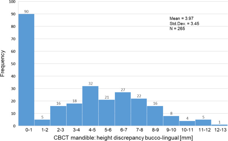
Results: The CBCT images of an unusual jaw contour were observed in 8 cases (2.4%) in the maxilla on the left and 10 cases (3%) in the maxilla on the right. In the mandible, a jaw contour deviates in 39 cases (12.1%) on the left side and 39 cases (12.1%) on the right side. A height difference was detected in the upper jaw in 307 cases and the lower jaw in 265 cases. The discrepancy was 2.09 mm (± 2.25 mm) in the maxilla and 3.97 mm (± 3.45 mm) in the mandible.
Conclusions: The CBCT scan provides useful information to avoid complications in the preoperative planning phase and surgical planning in implant dentistry.
Keywords: Alveolar ridge contour; Available bone width; Cone-beam computed tomography; Dental implant; Diagnosis/clinical assessment; Lingual undercuts; Submandibular fossa.
© 2023. The Author(s).
Conflict of interest statement
Ali Reza Ketabi, Andree Piwowarczyk, Matthias C. Schulz, Hans-Christoph Lauer and Stefan Hassfeld hereby declare that they have no conflict of interests.
Figures


Similar articles
-
Implant-guided volumetric analysis of edentulous maxillary bone with cone-beam computerized tomography scan. Maxillary sinus pneumatization classification.J Oral Implantol. 2012 Aug;38(4):377-90. doi: 10.1563/AAID-JOI-D-11-00212. J Oral Implantol. 2012. PMID: 22913308
-
Dental implant imaging: TeraRecon's Dental 3D Cone Beam Computed Tomography System.Dent Implantol Update. 2007 Jun;18(6):41-5. Dent Implantol Update. 2007. PMID: 17682685
-
The reliability of surgeons to avoid traumatic insertion of dental implants into high-risk regions: a panoramic radiograph study.BMC Oral Health. 2020 Apr 6;20(1):96. doi: 10.1186/s12903-020-01093-8. BMC Oral Health. 2020. PMID: 32252728 Free PMC article.
-
Radiographic modalities for diagnosis and treatment planning in implant dentistry.Implant Soc. 1995;5(5):7-11. Implant Soc. 1995. PMID: 9571835 Review.
-
Simultaneous implant placement and bone grafting with particulate mineralized allograft in sites with buccal wall defects, a three-year follow-up and review of literature.J Craniomaxillofac Surg. 2014 Jul;42(5):552-9. doi: 10.1016/j.jcms.2013.07.026. Epub 2013 Aug 22. J Craniomaxillofac Surg. 2014. PMID: 24529349 Review.
Cited by
-
The Clinical Efficacy and Safety of ErhBMP-2/BioCaP/β-TCP as a Novel Bone Substitute Using the Tooth-Extraction-Socket-Healing Model: A Proof-of-Concept Randomized Controlled Trial.J Clin Periodontol. 2025 Feb;52(2):299-309. doi: 10.1111/jcpe.14084. Epub 2024 Oct 31. J Clin Periodontol. 2025. PMID: 39478364 Free PMC article. Clinical Trial.
References
Publication types
MeSH terms
Substances
LinkOut - more resources
Full Text Sources
