. 2023 Jun 27;7(12):2681-2693.
doi: 10.1182/bloodadvances.2022008673.

Proteome alterations in erythrocytes with PIEZO1 gain-of-function mutations

Affiliations

Proteome alterations in erythrocytes with PIEZO1 gain-of-function mutations

Immacolata Andolfo et al. Blood Adv. .

Abstract

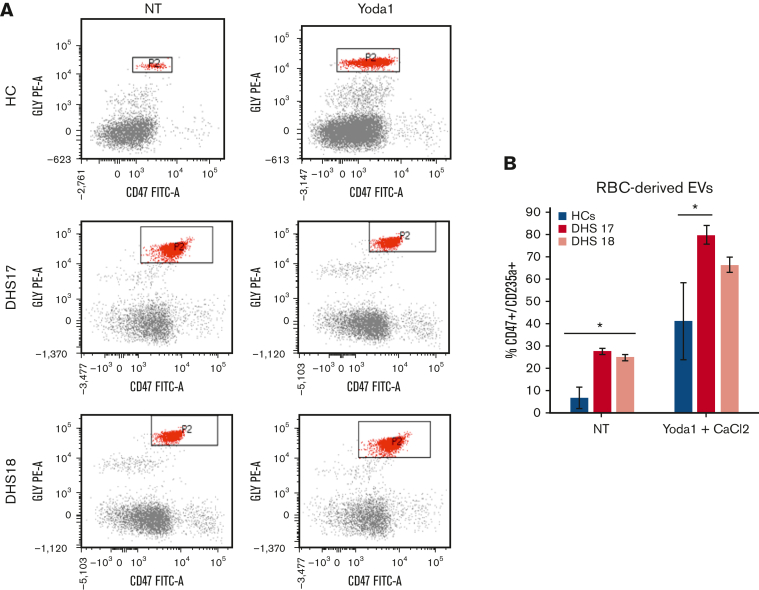
Gain-of-function mutations in PIEZO1 cause dehydrated hereditary stomatocytosis (DHS) or hereditary xerocytosis, an autosomal dominant hemolytic anemia characterized by high reticulocyte count, a tendency to macrocytosis, and mild jaundice, as well as by other variably penetrant clinical features, such as perinatal edema, severe thromboembolic complications after splenectomy, and hepatic iron overload. PIEZO1 mutations in DHS lead to slowed inactivation kinetics of the ion channel and/or facilitation of channel opening in response to physiological stimuli. To characterize the alterations of red blood cell proteome in patients with mutated PIEZO1, we used a differential approach to compare the proteome of patients with DHS (16 patients from 13 unrelated ancestries) vs healthy individuals. We identified new components in the regulation of the complex landscape of erythrocytes ion and volume balance mediated by PIEZO1. Specifically, the main impaired processes in patients with DHS were ion homeostasis, transmembrane transport, regulation of vesicle-mediated transport, and the proteasomal catabolic process. Functional assays demonstrated coexpression of PIEZO1 and band 3 when PIEZO1 was activated. Moreover, the alteration of the vesicle-mediated transport was functionally demonstrated by an increased vesiculation rate in patients with DHS compared with healthy controls. This finding also provides an explanation of the pathogenetic mechanism underlying the increased thrombotic rate observed in these patients. Finally, the newly identified proteins, involved in the intracellular signaling pathways altered by PIEZO1 mutations, could be used in the future as potential druggable targets in DHS.


Conflict of interest statement

Conflict-of-interest disclosure: The authors declare no competing financial interests.

Figures

None
Graphical abstract
Figure 1.
Figure 1.
Differentially expressed proteins (clustered for PIEZO1 protein domains) in patients with DHS compared with HCs. (A) Cytoscape graphical representation of proteins identified as differentially expressed in proteomic analyses, and clustered based on their identification within patient samples carrying a mutation in specific domains of PIEZO1. Red boxes refer to proteins identified in patients heterozygous and homozygous for mutations in THU7-THU8 domains; half red and yellow boxes indicate proteins solely shared between patients heterozygous and homozygous for mutation in THU7-THU8 domains. Purple, dark green, orange, and light green boxes refer to proteins specifically identified in patients that are heterozygous carrying mutations in CAP, CLASP, ANCHOR, and THU4-5 domains, respectively. Light-blue boxes refer to proteins differentially expressed and shared between at least 2 different conditions. (B) ClueGO functional analysis representation of biological processes including all differentially expressed proteins. GO was used as database; a P value Benjamini-Hochberg correction cutoff of 0.05 was applied. Cluster 1 in pink and cluster 2 in blue represent upregulated and downregulated, respectively. Node sizes were correlated with the specific P value.
Figure 1.
Figure 1.
Differentially expressed proteins (clustered for PIEZO1 protein domains) in patients with DHS compared with HCs. (A) Cytoscape graphical representation of proteins identified as differentially expressed in proteomic analyses, and clustered based on their identification within patient samples carrying a mutation in specific domains of PIEZO1. Red boxes refer to proteins identified in patients heterozygous and homozygous for mutations in THU7-THU8 domains; half red and yellow boxes indicate proteins solely shared between patients heterozygous and homozygous for mutation in THU7-THU8 domains. Purple, dark green, orange, and light green boxes refer to proteins specifically identified in patients that are heterozygous carrying mutations in CAP, CLASP, ANCHOR, and THU4-5 domains, respectively. Light-blue boxes refer to proteins differentially expressed and shared between at least 2 different conditions. (B) ClueGO functional analysis representation of biological processes including all differentially expressed proteins. GO was used as database; a P value Benjamini-Hochberg correction cutoff of 0.05 was applied. Cluster 1 in pink and cluster 2 in blue represent upregulated and downregulated, respectively. Node sizes were correlated with the specific P value.
Figure 2.
Figure 2.
Validation analysis of the transport, vesicle, and endocytosis pathways by multiple-reaction monitoring analysis. (A) Graphic representation obtained by the GraphPad software of the log2 normalized mean areas for all the proteins validated in patients with DHS (red boxes) compared with HCs (blue boxes). (B) Left: expression levels of OSR1 protein in patients with DHS with mutations in the mechanosensing module (green boxes) compared with patients with transduction module/ion-conducting pore mutations (purple boxes). Right: phosphorylation state analysis of OSR1 peptide (from amino acid 185-202) phosphorylated and nonphosphorylated in patients with DHS with mutations in the mechanosensing module (green boxes) compared with patients with transduction module/ion-conducting pore mutations (purple boxes). The representations were obtained by GraphPad software. A 2-way ANOVA test in a multiple comparison way was applied; ∗P ≤ .05; ∗∗P ≤ .01; ∗∗∗P ≤ .001; and ∗∗∗∗P ≤ .0001.
Figure 3.
Figure 3.
Increased levels of RBC-derived EVs in patients with PIEZO1 GoF mutations. (A) Flow cytometry analysis for RBC-derived EVs. Glycophorin A or CD235a, y-axis, and CD47 (x-axis) (axis values in log scale). Window P2 has CD235a+/Cd47+ cell populations that represent RBC-derived EVs. The analysis was performed in blood samples from HCs and 2 patients with DHS with PIEZO1 mutations (DHS17 and DHS18), not treated or treated with Yoda1 (PIEZO1 activator) and CaCl2. (B) RBC-derived EVs numbers in samples from flow cytometry analysis shown in panel A.
Figure 4.
Figure 4.
Band 3 and ABCB6 overexpression in plasma membrane protein of RBCs from patients with DHS and PIEZO1–band 3 colocalization. (A) Left: representative immunoblot of RBC membrane proteins showing band 3 and ABCB6 protein expression normalized to β-actin in the patients DHS1, DHS2, DHS3, DHS4, DHS5, and DHS6 compared with HCs (2 pools of HCs, each of n = 3). Right: densitometric analysis of 3 representative western blotting is shown for band 3 and ABCB6 proteins. Data are presented as mean ± standard deviation; ∗P < .05; ∗∗P < .01 (Student t test). (B) Representative confocal imaging by ZEISS LSM 980 Airyscan 2 of HC RBCs treated with vehicle (upper panel) compared with HC RBCs treated with 2.5 μM Yoda1 (10 minutes) (middle panel) and HC RBCs treated with 2.5 μM Yoda1 (20 minutes) (lower panel). Rabbit anti-PIEZO1 antibody is shown in green, mouse anti–band 3 is shown in red. The overlapping of both signals (MERGE) is shown on the right (yellow). (C) Quantification by Pearson correlation of PIEZO1–band 3 colocalization in untreated HC RBCs, HC RBCs + 2.5 μM Yoda1 (10 minutes), and HC RBCs + 2.5 μM Yoda1 (20 minutes) is shown. Data shown are the means of 12 independent acquisitions ± standard error of the mean; P < .05 for trend (ANOVA test), ∗P < .05 (post hoc correction by Tukey multiple comparisons test).

References

    1. Cahalan SM, Lukacs V, Ranade SS, Chien S, Bandell M, Patapoutian A. Piezo1 links mechanical forces to red blood cell volume. Elife. 2015;4 - PMC - PubMed
    1. Jiang F, Yin K, Wu K, et al. The mechanosensitive Piezo1 channel mediates heart mechano-chemo transduction. Nat Commun. 2021;12(1):869. - PMC - PubMed
    1. Li J, Hou B, Tumova S, et al. Piezo1 integration of vascular architecture with physiological force. Nature. 2014;515(7526):279–282. - PMC - PubMed
    1. Ranade SS, Qiu Z, Woo SH, et al. Piezo1, a mechanically activated ion channel, is required for vascular development in mice. Proc Natl Acad Sci U S A. 2014;111(28):10347–10352. - PMC - PubMed
    1. Rode B, Shi J, Endesh N, et al. Piezo1 channels sense whole body physical activity to reset cardiovascular homeostasis and enhance performance. Nat Commun. 2017;8(1):350. - PMC - PubMed

Publication types