. 2023 Jan 3;19(1):e1010563.
doi: 10.1371/journal.pgen.1010563. eCollection 2023 Jan.

ESR1 gene amplification and MAP3K mutations are selected during adjuvant endocrine therapies in relapsing Hormone Receptor-positive, HER2-negative breast cancer (HR+ HER2- BC)

Affiliations

ESR1 gene amplification and MAP3K mutations are selected during adjuvant endocrine therapies in relapsing Hormone Receptor-positive, HER2-negative breast cancer (HR+ HER2- BC)

Lorenzo Ferrando et al. PLoS Genet. .

Abstract

Background: Previous studies have provided a comprehensive picture of genomic alterations in primary and metastatic Hormone Receptor (HR)-positive, Human Epidermal growth factor Receptor 2 (HER2)-negative breast cancer (HR+ HER2- BC). However, the evolution of the genomic landscape of HR+ HER2- BC during adjuvant endocrine therapies (ETs) remains poorly investigated.

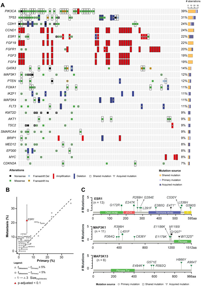
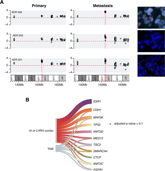
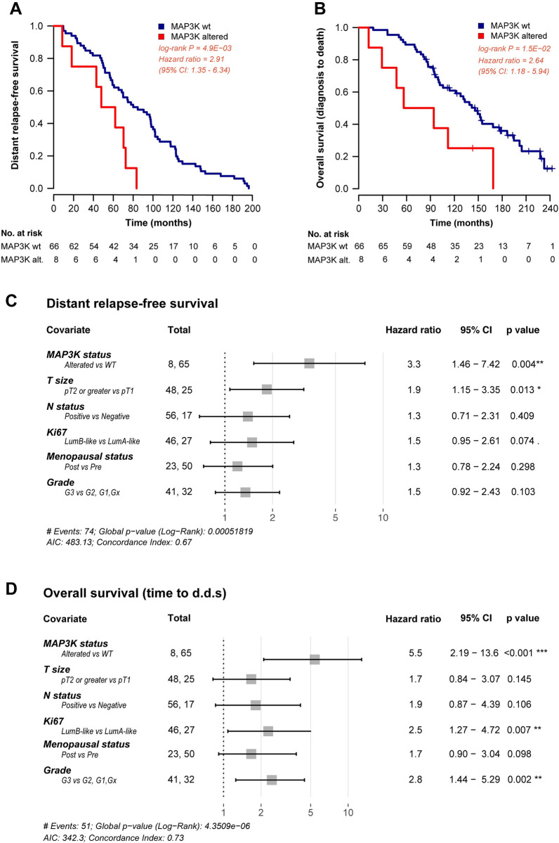
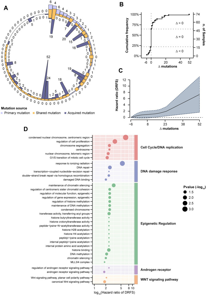
Methods and findings: We performed a genomic characterization of surgically resected HR+ HER2- BC patients relapsing during or at the completion of adjuvant ET. Using a customized panel, we comprehensively evaluated gene mutations and copy number variation (CNV) in paired primary and metastatic specimens. After retrieval and quality/quantity check of tumor specimens from an original cohort of 204 cases, 74 matched tumor samples were successfully evaluated for DNA mutations and CNV analysis. Along with previously reported genomic alterations, including PIK3CA, TP53, CDH1, GATA3 and ESR1 mutations/deletions, we found that ESR1 gene amplification (confirmed by FISH) and MAP3K mutations were enriched in metastatic lesions as compared to matched primary tumors. These alterations were exclusively found in patients treated with adjuvant aromatase inhibitors or LHRH analogs plus tamoxifen, but not in patients treated with tamoxifen alone. Patients with tumors bearing MAP3K mutations in metastatic lesions had significantly worse distant relapse-free survival (hazard ratio [HR] 3.4, 95% CI 1.52-7.70, p value 0.003) and worse overall survival (HR 5.2, 95% CI 2.10-12.8, p-value < 0.001) independently of other clinically relevant patient- and tumor-related variables.

Conclusions: ESR1 amplification and activating MAP3K mutations are potential drivers of acquired resistance to adjuvant ETs employing estrogen deprivation in HR+ HER2- BC. MAP3K mutations are associated with worse prognosis in patients with metastatic disease.


Conflict of interest statement

I have read the journal’s policy and the authors of this manuscript have the following competing interests: Giancarlo Pruneri reports honoraria from Novartis, Roche, Lilly and Exact Science. Gabriele Zoppoli reports travel grants from Novartis and Pfizer, and reagents for research from Citiva and ThermoFisher Scientific. Andrea Vingiani reports honoraria from Roche and Lilly. Marco Colleoni reports Research Grant from Roche. Elisabtta Munzone reports travel grants from Roche, Pfizer, Lilly and Novartis and reports receiving consultancy fees from Eisai, Exact Sciences, MSD Oncology, Daiichi Sankyo/Astra Zeneca, Pfizer and Seagen. Giuseppe Viale has received grants from Roche/Genentech and Astra Zeneca for his institution; consulting fees from Roche/Genentech, Astra Zeneca, MDS Oncology and Daiichi Sanyko; honoraria for lectures from Roche/Genentech, Astra Zeneca and Daiichi Sanyko; support for attending meetings from Roche/Genentech; and has served on Advisory Boards for Roche/Genentech, Astra Zeneca, Pfizer, MDS Oncology and Novartis. Lorenzo Ferrando, Anna Garuti, Claudio Vernieri, Antonino Belfiore, Luca Agnelli, Gianpaolo Dagrada, Diana Ivanoiu, Giuseppina Bonizzi, Luana Lippolis, Martina Dameri, Francesco Ravera, Luca Magnani and Alberto Ballestrero have no conflict of interest to disclose.

Figures

Fig 1
Fig 1. Repertoire of genomic alterations in primary and metastatic HR+ HER2- breast cancer (BC).
A) Recurrent driver somatic mutations and copy number variations identified in matched primary and metastatic HR+ HER2- BC specimens (n = 74) subjected to targeted sequencing (n = 148). Cases are shown in columns, whereas genes are shown in rows. Mutation types are color-coded according to the legend. The total number of alterations detected in individual genes is displayed on the bar plot (right). For graphical purpose, only the top 25 genes are shown. B) The scatter plot reports the mutational frequencies in matched primary (n = 74) and metastatic (n = 74) tumor samples. Color indicates statistical significance (p-value adjusted by false discovery rate ≤ 0.1), the shape of the points reflects the difference between mutational frequency in metastatic vs. primary tumor samples (triangle = frequency difference ≥ 5%, circle = frequency difference < 5%). Confidence intervals for proportions is reported for significant genes. C) Schematic representation of the protein domains of ESR1, MAP3K1 and MAP3K13 and of the somatic mutations in matched primary and metastatic HR+ HER2- BC specimens (n  =  74). Mutations are color-coded according to the legend, and their overall occurrence is represented on the y-axis.
Fig 2
Fig 2. ESR1 enrichment in metastatic HR+ HER2- BC samples.
A) ESR1 gene sCNV in three representative cases. Each row represents a case, and each box indicates the log-ratio levels for matched primary and metastatic tumor specimens. Red square shows the exact ESR1 region. The red background highlights the amplification of the ESR1 gene. FISH-based validation of each ESR1 amplification is also shown (right panel). B) Recurrent genomic alterations in metastatic tumor specimens, and their association with different types of endocrine therapy (ET). ET is classified according to specific clinically relevant groups. Statistically significant associations are shown as stars (adjusted p-value = 0.1).
Fig 3
Fig 3. Association between MAP3K alterations and clinical outcome.
Kaplan Meier curves displaying distant relapse-free survival (DRFS) (A) and overall survival (OS) (B) in patients with MAP3K gene alteration (red curves) as compared with patients with wild-type MAP3K status (blue curves). Forest plots indicating the hazard ratios for DRFS (C) and OS (D), and the corresponding confidence intervals, in MAP3K-altered and MAP3K-wild type patients. Multivariable Cox analysis is adjusted for tumor size, lymph node involvement, Ki67, menopausal status and tumor grade of the primary tumor.
Fig 4
Fig 4. Genomic spectrum of acquired driver alterations.
A) The circle graph represents for each case (n = 74) the proportion of driver mutations detected in primary and/or metastatic tumor samples. Outer numbers represent mutations of eBC, inner numbers represent mutations of mBC. B) Cumulative frequency of the difference (Δ) between number of mutations in metastatic vs. primary tumor samples (Δ < 0, number of driver mutations in the primary tumor greater than in the corresponding metastatic sample; Δ = 0, equal number of driver mutations in primary vs. metastatic tumor; Δ > 0, number of driver mutations in the primary sample lower than in the metastatic sample. C) Non-linear relationship between the difference of driver mutations in metastasis/primary pair (Δ, x-axis), and DRFS hazard ratio of Schoenfeld residuals (y-axis). The analysis is adjusted for T/N status, Ki67, menopausal status and tumor grade. The solid line represents a penalized spline fit of the predicting variables, while the dashed lines show 95% confidence intervals. D) Functional analysis of Gene Ontology (GO) terms associated to cell cycle, DDR, epigenetic regulation, androgen receptor activity and WNT signaling pathway. The size of the dots is inversely proportional to the p values of estimated hazard ratio (x-axis) displayed in log10 scale. P values are reported in S5 Table.

References

    1. Lefebvre C, Bachelot T, Filleron T, Pedrero M, Campone M, Soria J-C, et al. Mutational Profile of Metastatic Breast Cancers: A Retrospective Analysis. PLoS Med. 2016. Dec;13(12):e1002201. doi: 10.1371/journal.pmed.1002201 - DOI - PMC - PubMed
    1. Yates LR, Knappskog S, Wedge D, Farmery JHR, Gonzalez S, Martincorena I, et al. Genomic Evolution of Breast Cancer Metastasis and Relapse. Cancer Cell. 2017. Aug 14;32(2):169–184.e7. doi: 10.1016/j.ccell.2017.07.005 - DOI - PMC - PubMed
    1. Razavi P, Chang MT, Xu G, Bandlamudi C, Ross DS, Vasan N, et al. The Genomic Landscape of Endocrine-Resistant Advanced Breast Cancers. Cancer Cell. 2018. Sep 10;34(3):427–438.e6. doi: 10.1016/j.ccell.2018.08.008 - DOI - PMC - PubMed
    1. Aftimos P, Oliveira M, Irrthum A, Fumagalli D, Sotiriou C, Gal-Yam EN, et al. Genomic and Transcriptomic Analyses of Breast Cancer Primaries and Matched Metastases in AURORA, the Breast International Group (BIG) Molecular Screening Initiative. Cancer Discov. 2021. Nov;11(11):2796–811. doi: 10.1158/2159-8290.CD-20-1647 - DOI - PMC - PubMed
    1. Jeselsohn R, Buchwalter G, De Angelis C, Brown M, Schiff R. ESR1 mutations—a mechanism for acquired endocrine resistance in breast cancer. Nat Rev Clin Oncol. 2015. Oct;12(10):573–83. - PMC - PubMed

Publication types