. 2023 Jan 10;120(2):e2204134120.
doi: 10.1073/pnas.2204134120. Epub 2023 Jan 3.

Dry eye disease in mice activates adaptive corneal epithelial regeneration distinct from constitutive renewal in homeostasis

Affiliations

Dry eye disease in mice activates adaptive corneal epithelial regeneration distinct from constitutive renewal in homeostasis

Joseph B Lin et al. Proc Natl Acad Sci U S A. .

Abstract

Many epithelial compartments undergo constitutive renewal in homeostasis but activate unique regenerative responses following injury. The clear corneal epithelium is crucial for vision and is renewed from limbal stem cells (LSCs). Using single-cell RNA sequencing, we profiled the mouse corneal epithelium in homeostasis, aging, diabetes, and dry eye disease (DED), where tear deficiency predisposes the cornea to recurrent injury. In homeostasis, we capture the transcriptional states that accomplish continuous tissue turnover. We leverage our dataset to identify candidate genes and gene networks that characterize key stages across homeostatic renewal, including markers for LSCs. In aging and diabetes, there were only mild changes with <15 dysregulated genes. The constitutive cell types that accomplish homeostatic renewal were conserved in DED but were associated with activation of cell states that comprise "adaptive regeneration." We provide global markers that distinguish cell types in homeostatic renewal vs. adaptive regeneration and markers that specifically define DED-elicited proliferating and differentiating cell types. We validate that expression of SPARC, a marker of adaptive regeneration, is also induced in corneal epithelial wound healing and accelerates wound closure in a corneal epithelial cell scratch assay. Finally, we propose a classification system for LSC markers based on their expression fidelity in homeostasis and disease. This transcriptional dissection uncovers the dramatically altered transcriptional landscape of the corneal epithelium in DED, providing a framework and atlas for future study of these ocular surface stem cells in health and disease.

Keywords: cornea; dry eye; epithelium; limbal; stem cell.


Conflict of interest statement

The authors declare no competing interest.

Figures
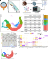
Fig. 1.
Fig. 1.
Single-cell atlas of the homeostatic mouse corneal epithelium. (A) Schematic for canonical model of LSC differentiation (created in BioRender). (B) Summary of experimental strategy (created in BioRender). (C) UMAP plot of scRNAseq atlas of the mouse corneal epithelium with table delineating cell frequencies for each population (n = 3 independent sequencing experiments). LSC, limbal stem cell; TAC, transit amplifying cell; Squam, squamous; Conj, conjunctiva. (D) Stacked bar plot showing relative frequencies for each cell cluster separated by sample. Colors correspond to those in C. (E) Schematic of cell adhesion molecules defining basal, wing, and squamous cell layers (created in BioRender). (F) Dot plot showing marker gene expression patterns. (G) UMAP plot showing cell cycle position assigned for each individual cell.
Fig. 2.
Fig. 2.
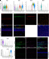
Markers for cell types in constitutive corneal epithelial renewal. (A) Violin plots showing expression of Gja1, Dsc2, Muc4, and Mki67. (B and C) Immunostaining of GJA1, DSC2, and MUC4 in the central cornea. Images reflect maximum projection across the entire thickness of the tissue section. (D) Ki67 immunostaining in the whole-mount cornea. This image was created by maximum projection of a Z-stack including only the basal layer of epithelial cells. (E) Violin plots showing expression of Ifitm3 and Krt19. (F) Immunostaining of the cornea for IFITM3 and K19. (Scale bars are the same for images in B and C.)
Fig. 3.
Fig. 3.
Gene regulatory networks characterizing corneal epithelial cell types in homeostasis. (A) Heat map depicting GRN activity in each cell cluster. Each row is a single GRN, and hierarchical clustering of cell types was performed based off of GRN activity. (B) UMAP plots of GRN activity for transcription factors that may define each stage of differentiation. (C) Heat maps show average expression of transcription factors and their target genes for each cell cluster.
Fig. 4.
Fig. 4.
Single-cell atlas of the corneal epithelium in a mouse model of DED. (A) Schematic of experimental approach (created in BioRender). (B) Fluorescein staining of ocular surface in control vs. DED at 7 d after lacrimal gland excision. The magenta dashed outline circumscribes areas of epithelial injury. (C) UMAP plots of corneal epithelial cell populations in control and DED with table delineating cell cluster frequencies. LSC, limbal stem cell; TAC, transit amplifying cell; Squam, squamous; Conj, conjunctiva; * denotes cell types elicited in DED. (D) Dot plot showing marker gene expression patterns. (E) UMAP plot of cell cycle positions assigned to each individual cell.
Fig. 5.
Fig. 5.
Transcriptional signature of adaptive corneal epithelial regeneration activated in DED. (A) Correlation matrix comparing transcriptomes of cell types in DED that are conserved from homeostasis. (B) Venn diagram showing the overlap of genes dysregulated in DED for cell types conserved from homeostasis. (C) Scatterplots compare the expression of genes in cell populations that are present in both control and DED. Listed genes are significantly dysregulated (i.e., adjusted P value < 0.05; |log2FC| ≥ 1). Genes that are up-regulated are listed in red and down-regulated in blue. (D) Venn diagram showing the overlap of genes that define DED-elicited cell types compared with their constitutive counterparts (i.e., LSC* vs. LSC; TAC1* vs. TAC1; TAC2* vs. TAC2; wing* vs. wing; squamous* vs. squamous). (E) Heat map depicts the average expression of genes that broadly distinguish cell types of constitutive turnover in homeostasis vs. adaptive regeneration activated in DED. (F) Heat map depicting the average expression of genes that distinguish proliferating and differentiated cells in homeostasis vs. DED. (G) Heat map of average expression of LSC markers in constitutive and DED-elicited cell types.
Fig. 6.
Fig. 6.
Role of SPARC in corneal epithelial wound healing. (A) Immunostaining for SPARC and Ki67 at 1 d following mechanical debridement of the central corneal epithelium. These images are of flat-mounted corneas and are maximum projections of the entire corneal epithelium. (B) Quantification of mean SPARC fluorescence intensity. Bars indicate mean ± SEM, and each circle represents an individual eye. Statistical significance was assessed using the Mann–Whitney test. (C) Heat map showing target genes of SPARC in human corneal epithelial cells. (D) Immunostaining for SPARC and FN1 at 1 d following mechanical debridement of the central corneal epithelium. These images are of flat-mounted corneas and are maximum projections of the entire corneal epithelium. (Scale bar same as shown for Fig. 6A.) (E) Representative images of scratch assay performed with human corneal epithelial cells when treated with SPARC. Yellow dashed lines indicate the edge of the cell-free scratch area. (F) Quantification of the remaining cell-free area relative to each treatment group’s initial average wound size. Each bar is colored corresponding to the labels in Fig. 7E and indicates the mean ± SEM calculated for n = 11 to 13 total scratches across three independent experiments. Statistical significance was assessed using the Brown–Forsythe ANOVA test for each time point (24-h omnibus P value < 0.01). For the 24-h time point, we performed a post hoc Dunnett’s T3 multiple comparisons test for all pairwise comparisons. *P < 0.05 and **P < 0.01.

Similar articles

Cited by

References

    1. Di Girolamo N., Moving epithelia: Tracking the fate of mammalian limbal epithelial stem cells. Prog. Retin. Eye Res. 48, 203–225 (2015). - PubMed
    1. Chew H. F., Limbal stem cell disease: Treatment and advances in technology. Saudi J. Ophthalmol. 25, 213–218 (2011). - PMC - PubMed
    1. Schermer A., Galvin S., Sun T. T., Differentiation-related expression of a major 64K corneal keratin in vivo and in culture suggests limbal location of corneal epithelial stem cells. J. Cell Biol. 103, 49–62 (1986). - PMC - PubMed
    1. Thoft R. A., Friend J., The X, Y, Z hypothesis of corneal epithelial maintenance. Invest. Ophthalmol. Vis. Sci. 24, 1442–1443 (1983). - PubMed
    1. Altshuler A., et al. , Discrete limbal epithelial stem cell populations mediate corneal homeostasis and wound healing. Cell Stem Cell 28, 1248–1261.e8 (2021). - PMC - PubMed

Publication types