. 2022 Dec 16;3(4):101857.
doi: 10.1016/j.xpro.2022.101857. Epub 2022 Nov 16.

Non-invasive chimeric HaloTag labeling to study clustering and diffusion of membrane proteins

Affiliations

Non-invasive chimeric HaloTag labeling to study clustering and diffusion of membrane proteins

Yiran Chang et al. STAR Protoc. .

Abstract

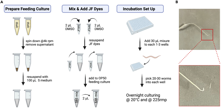
As live imaging plays an increasingly critical role in cell biology research, the desire to label and track individual protein molecules in vivo has been growing. To address this, in this protocol we describe steps for sparse labeling using two different HaloTag ligand dyes in C. elegans. This labeling approach is simple, is non-invasive, and preserves the view of the bulk protein population. We further describe how to carry out single-particle tracking experiments and extract information about particle diffusion behavior. For complete details on the use and execution of this protocol, please refer to Chang and Dickinson (2022).1.

Keywords: Biophysics; Cell Biology; Microscopy; Model Organisms; Single-molecule Assays.


Conflict of interest statement

Declaration of interests The authors declare no competing interests.

Figures

None
Graphical abstract
Figure 1
Figure 1
An overview of the study background and dual-labeling design, related to ‘before you begin’ (A) A cartoon illustration of how Halo works: the HaloTag binds covalently to its ligand dye. (B) Live images of PAR-3 segregation during C. elegans zygote polarization. Left panels: cortical Halo::PAR-3 labeled with JF646. Right panels: DIC channel images during polarization process. Scale bar: 10 μm. Figure modified from Chang and Dickinson. (C) TIRF images and cartoon illustrations of the dual-labeling experiment. Magenta represents far-red/abundant channel and green represents red/sparse channel. Scale bar: 10 μm. Figure modified from Chang and Dickinson.
Figure 2
Figure 2
Standard operating procedure for labeling the HaloTag in C. elegans, related to ‘preparing C. elegans for HaloTag imaging’ (A) Cartoon illustration of the procedure for JF dye feeding in C. elegans. Created with BioRender.com. (B) A hook shaped worm pick. The tip of the worm pick is zoomed out in the lower panel.
Figure 3
Figure 3
The workflow and interpretation of the MSD analysis, related to ‘Data Analysis’ (A) An illustration of the data analysis workflow. The appearance of the example data/results are shown to the right. Scale bar: 10 μm. (B) Correct and erroneous results from Utrack particle tracking. (a) Correct detection step result. (b) Erroneous detection step result: dimmer foci are not detected (white arrows). (c) Correct linking step result, orange lines represent the gaps in the tracks that have been connected by the software. (d) Erroneous linking step result: long straight lines and/or sharp turns (white arrows), and extensive gap filling (orange lines). (C) Graphical illustration of the interpretation of MSD plots and anomalous parameters. Figure modified from Chang and Dickinson. (D) An example illustration of the MSD analysis. Engineered Halo::PAR-3 clusters were labeled with JF585, data were pooled from 3 embryos. Left: the MSD curves. Blue lines are the MSD curve of each cluster tracked. Black line represents the averaged MSD curve. Middle: the averaged MSD curve. Right: the first derivative, or the slope, of the averaged MSD plot. Figure modified from Chang and Dickinson.

References

    1. Chang Y., Dickinson D.J. A particle size threshold governs diffusion and segregation of PAR-3 during cell polarization. Cell Rep. 2022;39:110652. doi: 10.1016/j.celrep.2022.110652. - DOI - PMC - PubMed
    1. Los G.V., Encell L.P., McDougall M.G., Hartzell D.D., Karassina N., Zimprich C., Wood M.G., Learish R., Ohana R.F., Urh M., et al. HaloTag: a novel protein labeling technology for cell imaging and protein analysis. ACS Chem. Biol. 2008;3:373–382. doi: 10.1021/cb800025k. - DOI - PubMed
    1. Grimm J.B., Muthusamy A.K., Liang Y., Brown T.A., Lemon W.C., Patel R., Lu R., Macklin J.J., Keller P.J., Ji N., Lavis L.D. A general method to fine-tune fluorophores for live-cell and in vivo imaging. Nat. Methods. 2017;14:987–994. doi: 10.1038/nmeth.4403. - DOI - PMC - PubMed
    1. Grimm J.B., English B.P., Chen J., Slaughter J.P., Zhang Z., Revyakin A., Patel R., Macklin J.J., Normanno D., Singer R.H., et al. A general method to improve fluorophores for live-cell and single-molecule microscopy. Nat. Methods. 2015;12:244–250. doi: 10.1038/nmeth.3256. 3 p following 250. - DOI - PMC - PubMed
    1. Grimm J.B., English B.P., Choi H., Muthusamy A.K., Mehl B.P., Dong P., Brown T.A., Lippincott-Schwartz J., Liu Z., Lionnet T., Lavis L.D. Bright photoactivatable fluorophores for single-molecule imaging. Nat. Methods. 2016;13:985–988. doi: 10.1038/nmeth.4034. - DOI - PubMed

Publication types