. 2022 Dec 16;3(4):101872.
doi: 10.1016/j.xpro.2022.101872. Epub 2022 Nov 7.

Protocol for SARS-CoV-2 infection of kidney organoids derived from human pluripotent stem cells

Affiliations

Protocol for SARS-CoV-2 infection of kidney organoids derived from human pluripotent stem cells

Elena Garreta et al. STAR Protoc. .

Abstract

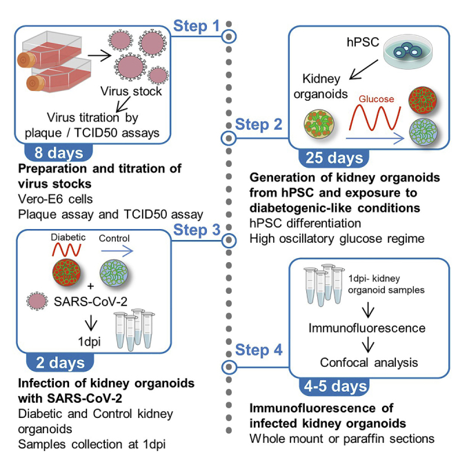
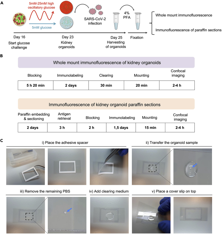
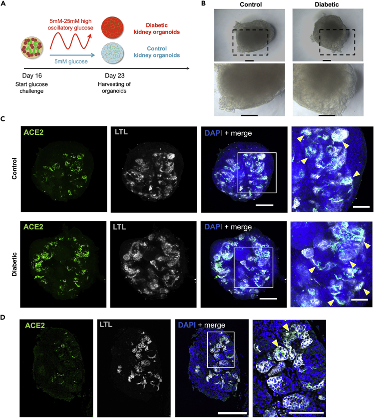
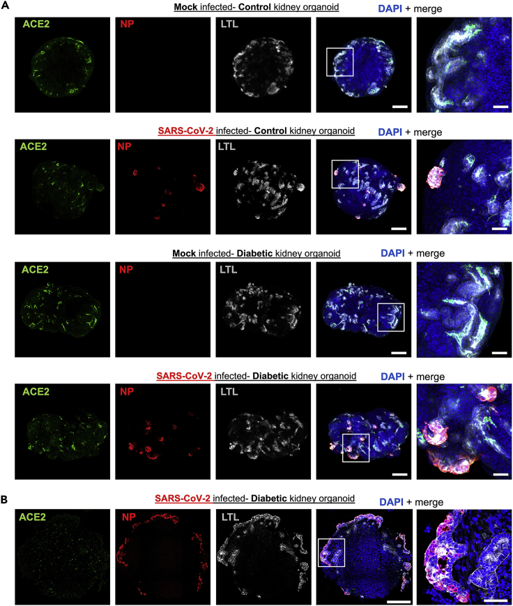
This protocol presents the use of SARS-CoV-2 isolates to infect human kidney organoids, enabling exploration of the impact of SARS-CoV-2 infection in a human multicellular in vitro system. We detail steps to generate kidney organoids from human pluripotent stem cells (hPSCs) and emulate a diabetic milieu via organoids exposure to diabetogenic-like cell culture conditions. We further describe preparation and titration steps of SARS-CoV-2 virus stocks, their subsequent use to infect the kidney organoids, and assessment of the infection via immunofluorescence. For complete details on the use and execution of this protocol, please refer to Garreta et al. (2022).1.

Keywords: Cell Differentiation; Cell culture; Microbiology; Microscopy; Organoids; Stem Cells.


Conflict of interest statement

Declaration of interests A patent has been submitted to use human organoids to study SARS-CoV-2 infections and possibly develop new therapies. J.M.P. is a shareholder of Apeiron Biologics, which is developing ACE2 decoys for COVID-19 therapy.

Figures

None
Graphical abstract
Figure 1
Figure 1
Typical morphology of hPSC colonies cultured on VTN-N-coated plates with E8 medium (A) Representative bright field images of the hiPSC line CBiPS1sv-4F-40 and the hESC line ES[4] at day 2, day 3 and day 5 after passaging. After 5 days in culture, hPSC colonies show nearly 80% confluency. (B) Representative bright field image taken 24 h after thawing an hPSC cryovial that shows dead floating hPSCs because of a failed thawing procedure. (C) Representative bright field image taken 48 h after hPSC passaging that shows very few small cell clusters indicating a failed passage procedure. (D) Representative bright field images of hPSC colonies with spontaneous differentiation. Yellow arrows indicate differentiated cells. Scale bars in (A-D), 100 μm.
Figure 2
Figure 2
Propagation and titration of SARS-CoV-2 virus in Vero-E6 Cells (A) Representative bright field image of Vero-E6 Cell monolayer with apparent CPE 72 h after SARS-CoV-2 virus inoculation. Scale bar, 100 μm. (B) Schematics of the virus serial dilutions preparation. (C) Schematics of a 6-well plate used for the plaque assay after staining with the crystal violet solution (left) and the corresponding image of a real plaque assay plate (right) showing the formation of the plaques. Note that the well corresponding to the 10-7 virus dilution does not show plaque formation but presents a scratch (indicated with a yellow circle) that has been accidentally performed when removing the agarose layer with the flat spoon and can be confounded with a plaque. (D) Representative fluorescent image of a typical 96-well plate prepared for virus titer assessment by the TCID50 assay (left). The table (right) shows a representative quantification of the number of positive infected wells that are used to calculate the virus titer by the Spearman-Karber method.
Figure 3
Figure 3
Generation of human kidney organoids from hPSC (A) Timeline of kidney organoid generation from hPSCs. PPS: posterior primitive streak; IM: intermediate mesoderm; NPC: nephron progenitor cell; RV: renal vesicle; ACT: activin A; Hep: heparin. (B) Representative bright field images showing morphological changes in the cell monolayer from day 0 to day 3 of differentiation. (C) Representative bright field image of the cell monolayer at day 4 of differentiation showing a very compact and uniform appearance. (D) Example of a failed formation of a day 4 cell monolayer. Yellow asterisks indicate areas of loose clusters of cells or empty areas. (E) Representative bright field image of a day 6 spheroid (2 days after intermediate mesoderm-committed cell aggregation into spheroids). At this stage, cell spheroid is characterized by a well-compacted round morphology. (F) Example of a failed cell aggregation in a day 6 spheroid. Note that edges are broken and disaggregated. (G) Representative bright field image of a day 9 organoid that begin to show the formation of RVs in the organoid edges. (H) Representative bright field image of a day 11 RV-stage organoid containing many RVs. (I) Example of a failed formation of RVs in a day 11 kidney organoid. (J) Representative bright field image of a day 16 kidney organoid containing multiple nephron-like structures. (K) Examples of inefficient formation of nephron-like structures within day 16 kidney organoids. Scale bars in (B–K), 100 μm.
Figure 4
Figure 4
Methodology to analyze kidney organoids by immunofluorescence (A) Timeline of the procedure to emulate a diabetic-like milieu in kidney organoids, infect them with SARS-CoV-2 virus and then harvest and fix them for performing immunofluorescence analysis. (B) Overview of the required steps to perform whole mount immunofluorescence or immunofluorescence in paraffin sections of kidney organoids. (C) Representative photographs to illustrate the procedure for mounting and clearing immunolabeled whole kidney organoid samples for imaging in a confocal microscope.
Figure 5
Figure 5
Immunofluorescence analysis of kidney organoids after exposure to diabetogenic-like culture conditions (A) Schematic overview of the procedure to induce control or diabetic kidney organoids. (B) Representative bright field images of control or diabetic kidney organoids. Scale bars, 100 μm. (C) Representative confocal images of control or diabetic kidney organoids analyzed by whole mount immunofluorescence for the detection of ACE2 (green), LTL (gray) and DAPI (blue). Scale bars, 250 μm, 100 μm (high magnification views). (D) Example of representative confocal images that show the detection of ACE2 (green), LTL (gray) and DAPI (blue) by immunofluorescence in an organoid paraffin section. Scale bars, 250 μm, 100 μm (high magnification view). Yellow arrowheads in (C-D) indicate examples of LTL positive proximal tubule-like structures with ACE2 expressing cells.
Figure 6
Figure 6
Immunofluorescence analysis of control and diabetic kidney organoids upon SARS-CoV-2 infection (A) Representative confocal images of mock or SARS-CoV-2 infected kidney organoids at 1 dpi for the detection of ACE2 (green), NP (red), LTL (gray) and DAPI (blue) by whole mount immunofluorescence. Scale bars, 200 μm, 50 μm (high magnification views). (B) Example of representative confocal images that show the detection of ACE2 (green), NP (red), LTL (gray) and DAPI (blue) by immunofluorescence in an organoid paraffin section. Scale bars, 200 μm, 50 μm (high magnification view).

References

    1. Garreta E., Prado P., Stanifer M.L., Monteil V., Marco A., Ullate-Agote A., Moya-Rull D., Vilas-Zornoza A., Tarantino C., Romero J.P., et al. A diabetic milieu increases ACE2 expression and cellular susceptibility to SARS-CoV-2 infections in human kidney organoids and patient cells. Cell Metab. 2022;34:857–873.e9. doi: 10.1016/j.cmet.2022.04.009. - DOI - PMC - PubMed
    1. Monteil V., Kwon H., Prado P., Hagelkrüys A., Wimmer R.A., Stahl M., Leopoldi A., Garreta E., Hurtado del Pozo C., Prosper F., et al. Inhibition of SARS-CoV-2 infections in engineered human tissues using clinical-grade soluble human ACE2. Cell. 2020;181:905–913.e7. doi: 10.1016/j.cell.2020.04.004. - DOI - PMC - PubMed
    1. Garreta E., Prado P., Tarantino C., Oria R., Fanlo L., Martí E., Zalvidea D., Trepat X., Roca-Cusachs P., Gavaldà-Navarro A., et al. Fine tuning the extracellular environment accelerates the derivation of kidney organoids from human pluripotent stem cells. Nat. Mater. 2019;18:397–405. doi: 10.1038/s41563-019-0287-6. - DOI - PMC - PubMed
    1. Dhillon P., Park J., Hurtado del Pozo C., Li L., Doke T., Huang S., Zhao J., Kang H.M., Shrestra R., Balzer M.S., et al. The nuclear receptor ESRRA protects from kidney disease by coupling metabolism and differentiation. Cell Metab. 2021;33:379–394.e8. doi: 10.1016/j.cmet.2020.11.011. - DOI - PMC - PubMed
    1. Eriksen A.Z., Møller R., Makovoz B., Uhl S.A., tenOever B.R., Blenkinsop T.A. SARS-CoV-2 infects human adult donor eyes and hESC-derived ocular epithelium. Cell Stem Cell. 2021;28:1205–1220.e7. doi: 10.1016/j.stem.2021.04.028. - DOI - PMC - PubMed

Publication types