. 2022 Dec 16;3(4):101898.
doi: 10.1016/j.xpro.2022.101898. Epub 2022 Dec 5.

Generation of human elongating multi-lineage organized cardiac gastruloids

Affiliations

Generation of human elongating multi-lineage organized cardiac gastruloids

Zachary T Olmsted et al. STAR Protoc. .

Abstract

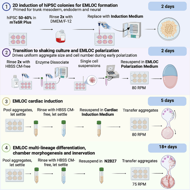
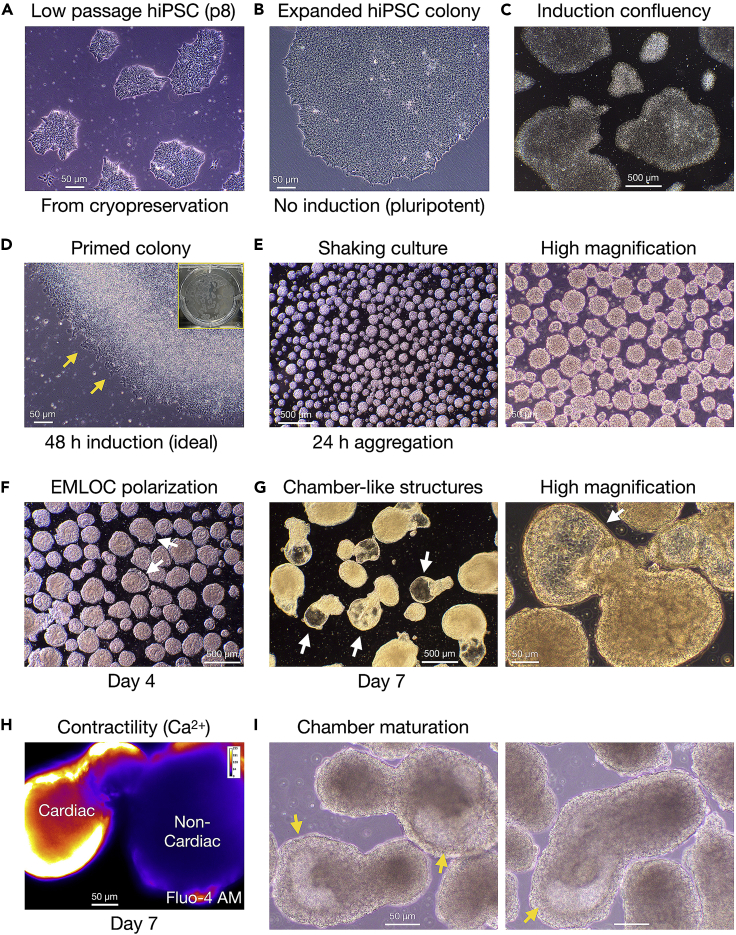
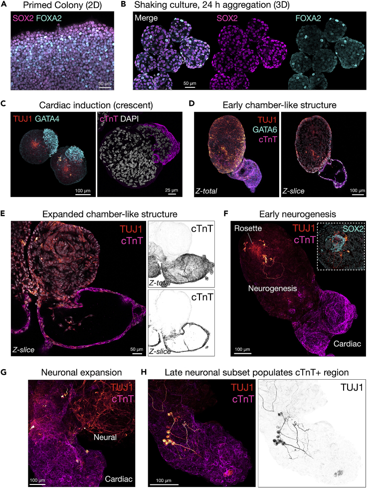
Human elongating multi-lineage organized (EMLOC) gastruloid technology captures key aspects of trunk neurodevelopment including neural integration with cardiogenesis. We generate multi-chambered, contractile EMLOC gastruloids with integrated central and peripheral neurons using defined culture conditions and signaling factors. hiPSC colonies are primed by activating FGF and Wnt signaling pathways for co-induced lineages. EMLOC gastruloids are then initialized with primed cells in suspension culture using timed exposure to FGF2, HGF, IGF1, and Y-27632. Cardiogenesis is stimulated by FGF2, VEGF, and ascorbic acid. For complete details on the use and execution of this protocol, please refer to Olmsted and Paluh (2022).1.

Keywords: Bioinformatics; Developmental biology; Neuroscience; Organoids; Stem Cells.


Conflict of interest statement

Declaration of interests Application for patent for which J.L.P. and Z.T.O. are co-inventors has been filed by the State University of New York Research Foundation (SUNYRF) with the US Patent Office on the EMLOC technology and detailed methods. U.S. patent filings: 63/311,498 and 63/419,507.

Figures

None
Graphical abstract
Figure 1
Figure 1
Overview of EMLOC formation and multi-lineage differentiation protocol Timeline of EMLOC formation and differentiation in four general stages: (1) 2D induction of hiPSC colonies, (2) transition to shaking culture and EMLOC early polarization, (3) EMLOC cardiac induction, and (4) EMLOC multi-lineage differentiation, chamber morphogenesis and innervation.
Figure 2
Figure 2
Workflow of EMLOC induction, polarization, and maturation stages (A) H3.1.1 low passage hiPSCs broken out on Matrigel after cryopreservation. (B) Expanded hiPSC colony in mTeSR Plus pluripotency medium. (C) Low magnification image of hiPSC cultures in mTeSR Plus at adequate confluency to begin 2D induction (∼60%). (D) Stage 1: Induction of 2D hiPSC colonies in Induction Medium (N2B27 basal medium with 3 μM CHIR 99021 and 40 ng/mL FGF2) at 48 h. By visual inspection, optimal colony induction is characterized by slight raised-edge character just as cells begin to migrate away from the colony border (yellow arrows). Inset is white light image of primed colonies just before generation of single cell suspensions. (E) Stage 2: Transition to shaking culture in EMLOC Polarization Medium. Single cell suspensions are generated and transferred to low-adhesion 6-well plates (2 × 10⁶ cells/well). Spontaneous aggregation (size range ∼50–100 μm) at 24 h. Low 5× magnification is shown (left) with high magnification image (right). (F) Stage 3: Early EMLOC polarization in Cardiac Induction Medium with cardiac crescent formation (white arrows) at day 4 post-aggregation. (G) Polarized EMLOCs with contractile chamber-like structures (white arrows) at low magnification (left) and high magnification (right). (H) Contracting cardiogenic region by live-cell calcium imaging using Fluo-4 AM dye in day 7 EMLOC. (I) Stage 4: maturation of EMLOC chamber-like structures (yellow arrows). Two fields are shown. Individual scale bars are provided for all images.
Figure 3
Figure 3
Polarization of biomarkers at key steps in EMLOC formation by immunofluorescence (A) Stage 1 (Induction Medium): immunofluorescence image of primed 2D hiPSC colony induced by CHIR 99021 and FGF2 in N2B27 basal medium. Co-expression of neuroectodermal biomarker SOX2 and mesendodermal biomarker FOXA2 is shown. (B) Stage 2 (EMLOC Polarization Medium): Individual spherical aggregates at 24 h post-aggregation in shaking culture. Expression pattern of SOX2 and FOXA2 is shown as merge (+DAPI) and separate channels. Seven aggregates are visible. (C) Stage 3 (Cardiac Induction Medium): day 4 EMLOCs with early polarized cardiac crescent (GATA4 left; cTnT right). Very early neurogenesis (TUJ1) is visible from single rosettes (left). (D) Early chamber-like structures (cTnT) and distinct neurogenesis compartment (TUJ1). EMLOC is co-stained with GATA6. Maximally projected Z-stack (Z-total) is shown with single Z-slice to highlight morphology. (E) Expanded chamber-like structure (cTnT) and distinct neurogenesis compartment (TUJ1). Z-total (top right) and single Z-slice (bottom right) of inverted cTnT channel is shown. (F) Stage 4 (N2B27, no supplements): early neurogenesis from single rosette (TUJ1) with maturing chamber-like structure (cTnT). Inset is high magnification image showing SOX2+ rosette. Early neurons are diverted away from the cardiac region. (G) Neuronal expansion (TUJ1) in neurogenesis compartment and early interaction with cTnT+ cardiac region. (H) A late neuronal subset (TUJ1) begins to populate the cTnT+ cardiac region. Inverted TUJ1 channel is shown (right). Individual scale bars are provided for all images.

References

    1. Olmsted Z.T., Paluh J.L. A combined human gastruloid model of cardiogenesis and neurogenesis. iScience. 2022;25:104486. - PMC - PubMed
    1. Chang E.A., Tomov M.L., Suhr S.T., Luo J., Olmsted Z.T., Paluh J.L., Cibelli J. Derivation of ethnically diverse human induced pluripotent stem cell lines. Sci. Rep. 2015;5:15234. - PMC - PubMed
    1. Tomov M.L., Olmsted Z.T., Dogan H., Gongorurler E., Tsompana M., Otu H.H., Buck M., Chang E.A., Cibelli J., Paluh J.L. Distinct and Shared Determinants of cardiomyocyte contractility in multi-lineage competent ethnically diverse human iPSCs. Sci. Rep. 2016;6:37637. - PMC - PubMed
    1. Gouti M., Tsakiridis A., Wymeersch F.J., Huang Y., Kleinjung J., Wilson V., Briscoe J. In vitro generation of neuromesodermal progenitors reveals distinct roles for wnt signaling in the specification of spinal cord and paraxial mesoderm identity. PLoS Biol. 2014;12:e1001937. - PMC - PubMed
    1. Olmsted Z.T., Stigliano C., Badri A., Zhang F., Williams A., Koffas M.A.G., Xie Y., Linhardt R.J., Cibelli J., Horner P.J., Paluh J.L. Fabrication of homotypic neural ribbons as a multiplex platform optimized for spinal cord delivery. Sci. Rep. 2020;10:12939. - PMC - PubMed

Publication types

LinkOut - more resources