. 2023 Jan 3;11(1):1.
doi: 10.1186/s40478-022-01485-7.

Differential vulnerability of the dentate gyrus to tauopathies in dementias

Affiliations

Differential vulnerability of the dentate gyrus to tauopathies in dementias

Allegra Kawles et al. Acta Neuropathol Commun. .

Abstract

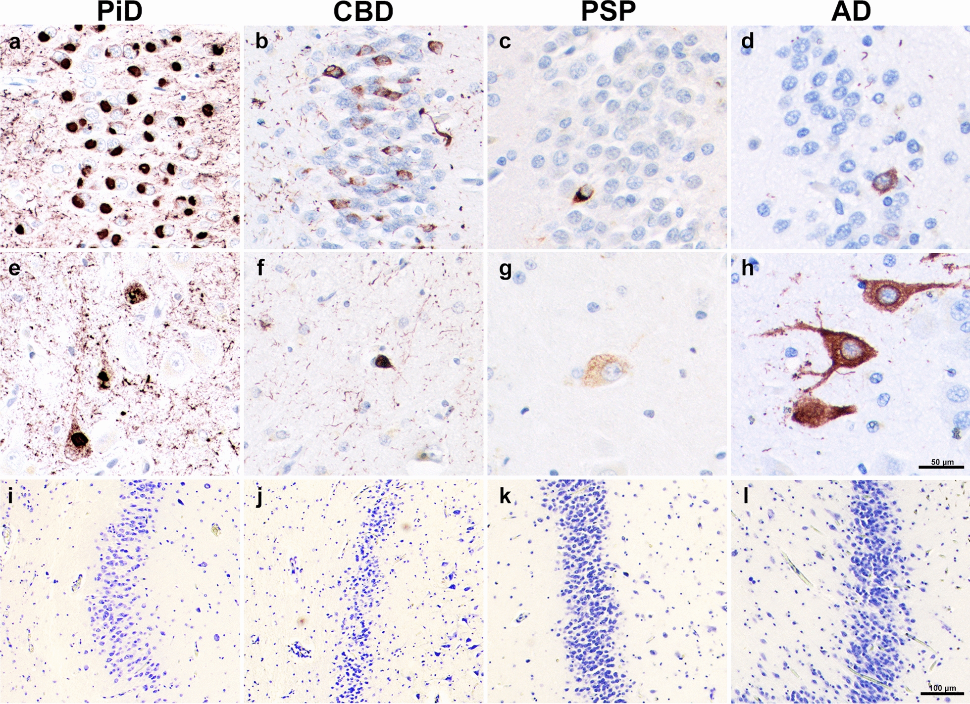
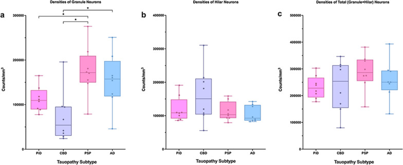
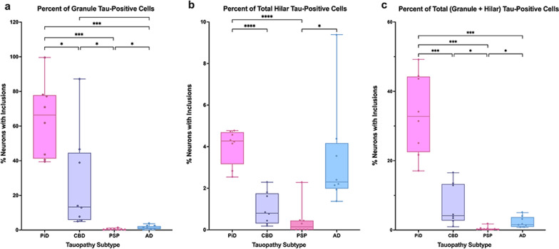
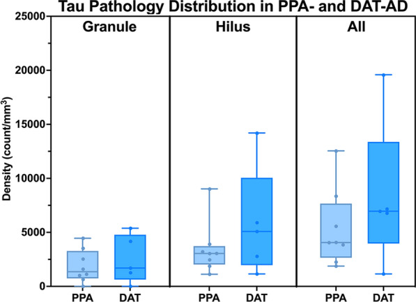
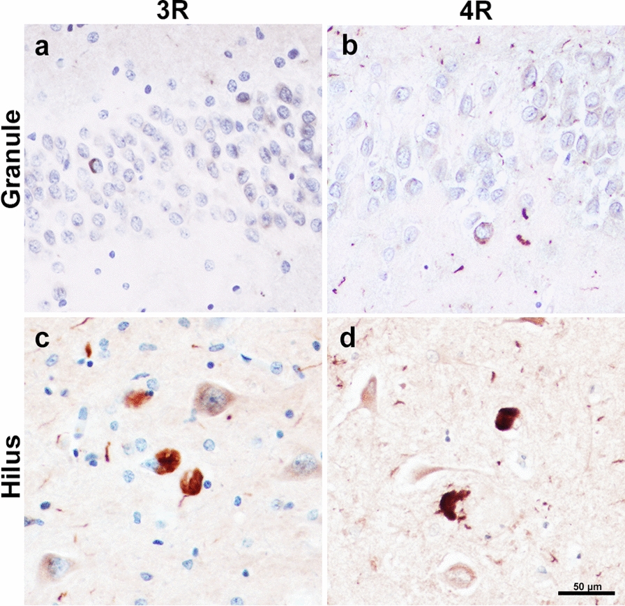
The dentate gyrus (DG), a key hippocampal subregion in memory processing, generally resists phosphorylated tau accumulation in the amnestic dementia of the Alzheimer's type due to Alzheimer's disease (DAT-AD), but less is known about the susceptibility of the DG to other tauopathies. Here, we report stereologic densities of total DG neurons and tau inclusions in thirty-two brains of human participants with autopsy-confirmed tauopathies with distinct isoform profiles-3R Pick's disease (PiD, N = 8), 4R corticobasal degeneration (CBD, N = 8), 4R progressive supranuclear palsy (PSP, N = 8), and 3/4R AD (N = 8). All participants were diagnosed during life with primary progressive aphasia (PPA), an aphasic clinical dementia syndrome characterized by progressive deterioration of language abilities with spared non-language cognitive abilities in early stages, except for five patients with DAT-AD as a comparison group. 51% of total participants were female. All specimens were stained immunohistochemically with AT8 to visualize tau pathology, and PPA cases were stained for Nissl substance to visualize neurons. Unbiased stereological analysis was performed in granule and hilar DG cells, and inclusion-to-neuron ratios were calculated. In the PPA group, PiD had highest mean total (granule + hilar) densities of DG tau pathology (p < 0.001), followed by CBD, AD, then PSP. PPA-AD cases showed more inclusions in hilar cells compared to granule cells, while the opposite was true in PiD and CBD. Inclusion-to-neuron ratios revealed, on average, 33% of all DG neurons in PiD cases contained a tau inclusion, compared to ~ 7% in CBD, 2% in AD, and 0.4% in PSP. There was no significant difference between DAT-AD and PPA-AD pathologic tau burden, suggesting that differences in DG burden are not specific to clinical phenotype. We conclude that the DG is differentially vulnerable to pathologic tau accumulation, raising intriguing questions about the structural integrity and functional significance of hippocampal circuits in neurodegenerative dementias.

Keywords: Dentate gyrus; Frontotemporal lobar degeneration; Primary progressive aphasia; Tauopathy.


Conflict of interest statement

The authors declare that they have no competing interests.

Figures

Fig. 1
Fig. 1
Tau-positive Pathology and Neurons in Aphasic Dementia. (a–d) Granule cell tau pathology visualized through AT8 immunohistochemical staining. (e–h) Hilar tau pathology revealed through AT8 immunohistochemical staining. (i–l) Granule neuronal density revealed through histochemical staining with cresyl violet. (a, e, i) was taken from case PiD 7, (b, f, j) was taken from CBD 6, (c, g, k) was taken from PSP 4, and (d, h, l) was taken from PPA-AD 3. PiD images show intense tau neuropathologic burden in both granule (a) and hilar (e) cells. CBD images show greater neuropathologic inclusions in granule cells (b), while PPA-AD has more inclusions in the hilar region (h). PSP shows uniformly sparse pathology (c, g). While PSP and AD show relatively preserved neuronal densities (k, l), there is no significant difference between neuronal densities across tauopathies. Scale bar = 50 µm in h, and applies to a–g. Scale bar = 100 µm in l, and applies to i–k
Fig. 2
Fig. 2
Densities of Dentate Gyrus Neurons in PPA due to Tauopathies. Box and whisker plots show densities of granule (a), hilar (b), and total (c) neurons in the dentate gyrus of PPA-PiD (N = 8), -CBD (N = 8), -PSP (N = 8), and -AD (N = 8) cases. PSP cases showed a significantly higher density of granule neurons compare to PiD and CBD (p < 0.05), and AD cases demonstrated a significantly higher density of granule neurons than CBD cases (p < 0.05). Total neuronal density was generally equal across all tauopathies. Whiskers indicate minimum and maximum densities, and the box delineates lower and upper quartiles. Each point represents the neuronal density of an individual case. Line within the box represents median
Fig. 3
Fig. 3
Percent of Dentate Gyrus Neurons affected by Tau-positive Inclusions in PPA. (a–c) Box and whisker plot show percent of neurons containing an inclusion in the dentate gyrus of PiD (N = 8), CBD (N = 8), PSP (N = 8), and AD (N = 8) cases, all with a clinical diagnosis of PPA. Percent of neurons containing an inclusion in granule (a), hilar (b), and all (c) dentate gyrus neurons was found by calculating an inclusion-to-neuron ratio and converting the quotient into a percent. Patterns of significance in ratio data closely followed those of inclusion data, where PiD showed a higher percent of tau-positive granule and hilar cells than all other groups. CBD showed a slight predilection for granule cell tau-positivity, and AD had a clear preference for tau-positivity in hilar regions. *p < 0.05; **p < 0.01; ***p < 0.001; ****p < 0.0001. Note the large difference in the relative scale of the y-axis across all bar graphs. Whiskers indicate minimum and maximum densities, and the box delineates lower and upper quartiles. Each dot represents an individual case. Line within the box represents median
Fig. 4
Fig. 4
Density of Tau Pathology in Amnestic (PPA) vs Aphasic (DAT) Dementia due to Alzheimer’s Disease. Box and whisker plot show the densities per cubic millimeter of neurofibrillary tau pathology of the AD type in participants diagnosed antemortem with primary progressive aphasia (PPA) (N = 8) or dementia of the Alzheimer’s type (DAT) (N = 5). Total DG counts (granule + hilar) are provided as well as stratified by granule and hilar cell populations. While DAT-AD had slightly more overall and hilar tau pathology, this difference did not reach significance. Whiskers indicate minimum and maximum densities, and the box delineates lower and upper quartiles. Each dot represents an individual PPA-AD or DAT-AD case. Line within the box represents median
Fig. 5
Fig. 5
3R- and 4R-tau-positive Inclusions in PPA due to Alzheimer’s Disease. 3R-positive pathology visualized through RD3 immunohistochemical staining in granule (a) and hilar cells (c); taken from case PPA-AD 7. 4R-positive pathology visualized through ET3 immunohistochemical staining in granule (b) and hilar cells (d); taken from case PPA-AD 5. Ratio of stereologic densities revealed ~ 1:1 ratio of 3R-to-4R immunopositivity in the granule cells, and ~ 1.5:1 ratio in hilar cells. Differences in 3R and 4R densities were not significantly different. Scale bar = 50 µm in d, and applies to a–c

References

    1. Langston RF, Stevenson CH, Wilson CL, Saunders I, Wood ER. The role of hippocampal subregions in memory for stimulus associations. Behav Brain Res. 2010;215:275–291. doi: 10.1016/j.bbr.2010.07.006. - DOI - PubMed
    1. Seok JW, Cheong C. Functional dissociation of hippocampal subregions corresponding to memory types and stages. J Physiol Anthropol. 2020;39:15. doi: 10.1186/s40101-020-00225-x. - DOI - PMC - PubMed
    1. Braak H, Braak E. Staging of Alzheimer's disease-related neurofibrillary changes. Neurobiol Aging. 1995;16:271–278. doi: 10.1016/0197-4580(95)00021-6. - DOI - PubMed
    1. Burke SN, Barnes CA. Senescent synapses and hippocampal circuit dynamics. Trends Neurosci. 2010;33:153–161. doi: 10.1016/j.tins.2009.12.003. - DOI - PMC - PubMed
    1. Morrison JH, Hof PR. Selective vulnerability of corticocortical and hippocampal circuits in aging and Alzheimer's disease. Prog Brain Res. 2002;136:467–486. doi: 10.1016/s0079-6123(02)36039-4. - DOI - PubMed

Publication types