

Rapid remodeling observed at mid-term in-vivo study of a smart reinforced acellular vascular graft implanted on a rat model
- PMID: 36597162
- PMCID: PMC9810246
- DOI: 10.1186/s13036-022-00313-9
Rapid remodeling observed at mid-term in-vivo study of a smart reinforced acellular vascular graft implanted on a rat model
Abstract
Background: The poor performance of conventional techniques used in cardiovascular disease patients requiring hemodialysis or arterial bypass grafting has prompted tissue engineers to search for clinically appropriate off-the-shelf vascular grafts. Most patients with cardiovascular disease lack suitable autologous tissue because of age or previous surgery. Commercially available vascular grafts with diameters of < 5 mm often fail because of thrombosis and intimal hyperplasia.
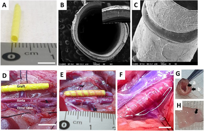
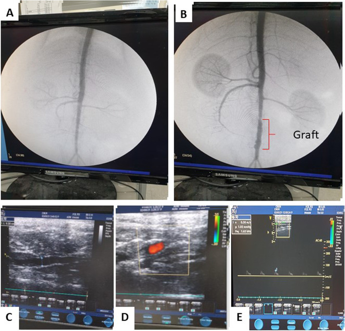
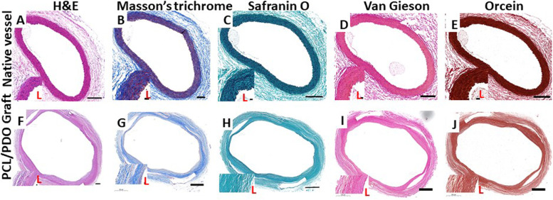
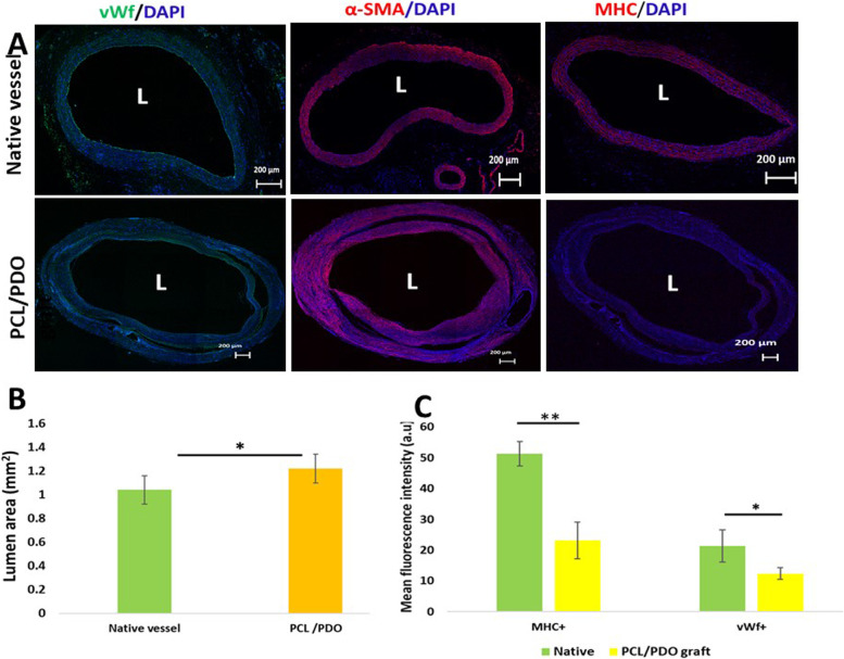
Result: Here, we tested tubular biodegradable poly-e-caprolactone/polydioxanone (PCL/PDO) electrospun vascular grafts in a rat model of aortic interposition for up to 12 weeks. The grafts demonstrated excellent patency (100%) confirmed by Doppler Ultrasound, resisted aneurysmal dilation and intimal hyperplasia, and yielded neoarteries largely free of foreign materials. At 12 weeks, the grafts resembled native arteries with confluent endothelium, synchronous pulsation, a contractile smooth muscle layer, and co-expression of various extracellular matrix components (elastin, collagen, and glycosaminoglycan).
Conclusions: The structural and functional properties comparable to native vessels observed in the neoartery indicate their potential application as an alternative for the replacement of damaged small-diameter grafts. This synthetic off-the-shelf device may be suitable for patients without autologous vessels. However, for clinical application of these grafts, long-term studies (> 1.5 years) in large animals with a vasculature similar to humans are needed.
Keywords: 3D printing; Electrospinning; Medium-term performance; Nanofibers; Rat abdominal aorta replacement model; Tissue regeneration; Vascular graft.
© 2022. The Author(s).
Conflict of interest statement
There is no competing interest.
Figures


Similar articles
-
Appropriate density of PCL nano-fiber sheath promoted muscular remodeling of PGS/PCL grafts in arterial circulation.Biomaterials. 2016 May;88:34-47. doi: 10.1016/j.biomaterials.2016.02.026. Epub 2016 Feb 23. Biomaterials. 2016. PMID: 26943048
-
A Comparative Study of an Anti-Thrombotic Small-Diameter Vascular Graft with Commercially Available e-PTFE Graft in a Porcine Carotid Model.Tissue Eng Regen Med. 2022 Jun;19(3):537-551. doi: 10.1007/s13770-021-00422-4. Epub 2022 Feb 15. Tissue Eng Regen Med. 2022. PMID: 35167044 Free PMC article.
-
Fast-degrading elastomer enables rapid remodeling of a cell-free synthetic graft into a neoartery.Nat Med. 2012 Jul;18(7):1148-53. doi: 10.1038/nm.2821. Nat Med. 2012. PMID: 22729285 Free PMC article.
-
Considerations in the Development of Small-Diameter Vascular Graft as an Alternative for Bypass and Reconstructive Surgeries: A Review.Cardiovasc Eng Technol. 2020 Oct;11(5):495-521. doi: 10.1007/s13239-020-00482-y. Epub 2020 Aug 18. Cardiovasc Eng Technol. 2020. PMID: 32812139 Review.
-
Polyglycerol sebacate-based elastomeric materials for arterial regeneration.J Biomed Mater Res A. 2024 Apr;112(4):574-585. doi: 10.1002/jbm.a.37583. Epub 2023 Jun 22. J Biomed Mater Res A. 2024. PMID: 37345954 Review.
Cited by
-
Vascular Damage and Repair - Are Small-Diameter Vascular Grafts Still the "Holy Grail" of Tissue Engineering?Physiol Res. 2024 May 31;73(Suppl 1):S335-S363. doi: 10.33549/physiolres.935294. Epub 2024 May 31. Physiol Res. 2024. PMID: 38836460 Free PMC article. Review.
-
Normal Blood Flow in Rat Abdominal Aorta: An Ultrasound Study.Biomedicines. 2025 Jun 5;13(6):1385. doi: 10.3390/biomedicines13061385. Biomedicines. 2025. PMID: 40564103 Free PMC article.
References
-
- Team. S. Heart disease statistics 2021. News; 2021.
LinkOut - more resources
Full Text Sources
