

MiR-138-5p suppresses the progression of lung cancer by targeting SNIP1
- PMID: 36597175
- PMCID: PMC9968603
- DOI: 10.1111/1759-7714.14791
MiR-138-5p suppresses the progression of lung cancer by targeting SNIP1
Abstract
Background: MicroRNAs (miRNAs) play crucial roles in the development of various cancers. Here, we aimed to evaluate the roles of miR-138-5p in lung cancer progression and the value of miR-138-5p in lung cancer diagnosis.
Methods: Quantitative real-time PCR was performed to examine the expressions of miR-138-5p and smad nuclear interacting protein 1 (SNIP1) mRNA. The diagnostic value of miR-138-5p was analyzed using receiver operating characteristic (ROC) curve analysis, sensitivity, and specificity. We explored the effect of miR-138-5p on cell proliferation and metastasis by CCK-8, colony formation, wound healing and transwell assays. Western blot was employed to detect the protein expression of SNIP1 and related genes. Lung cancer cell growth was evaluated in vivo using xenograft tumor assay.
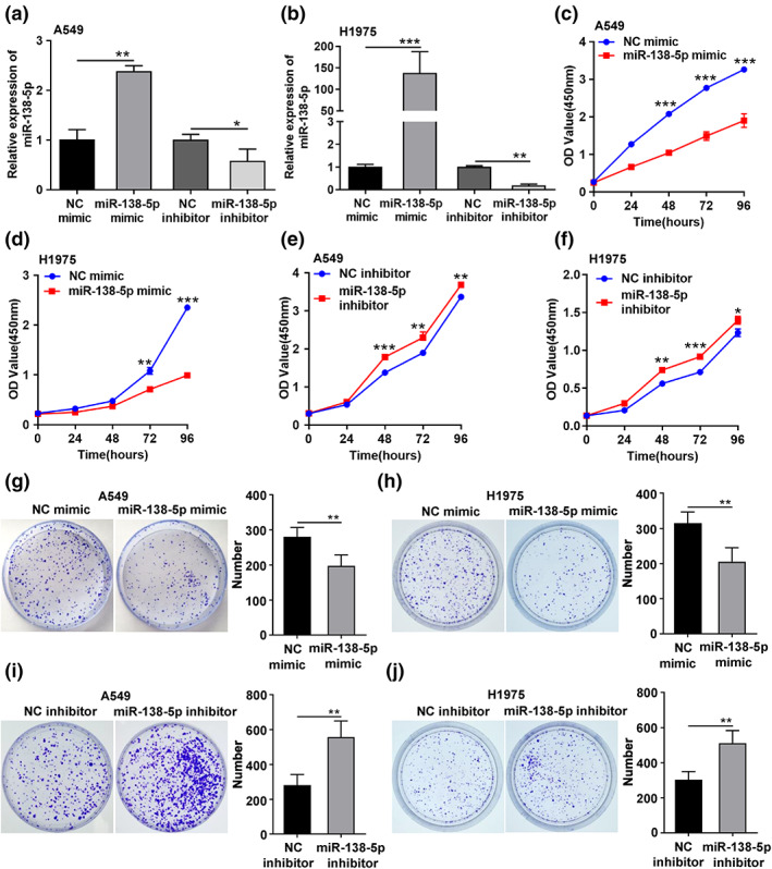
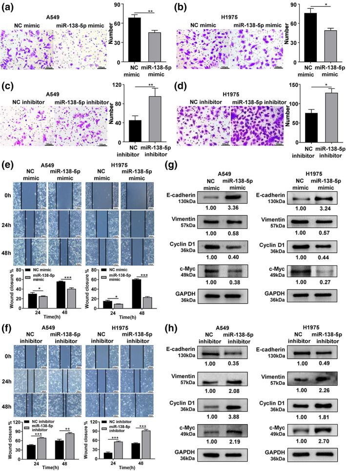
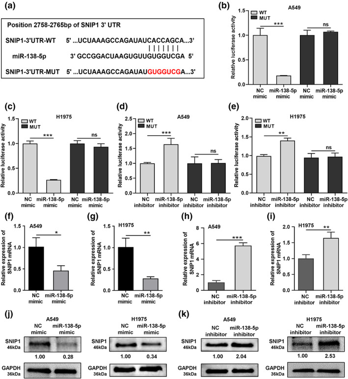
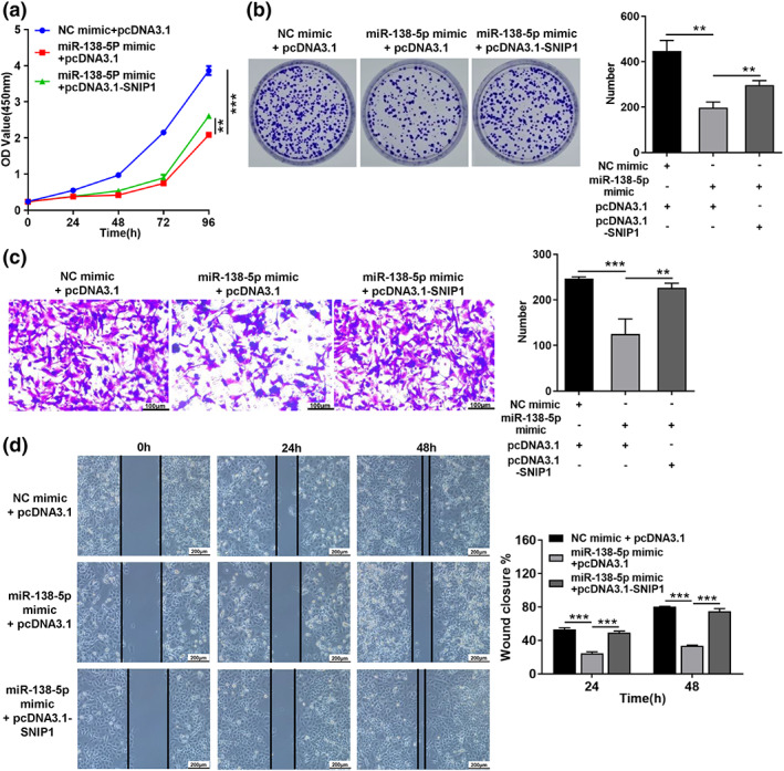
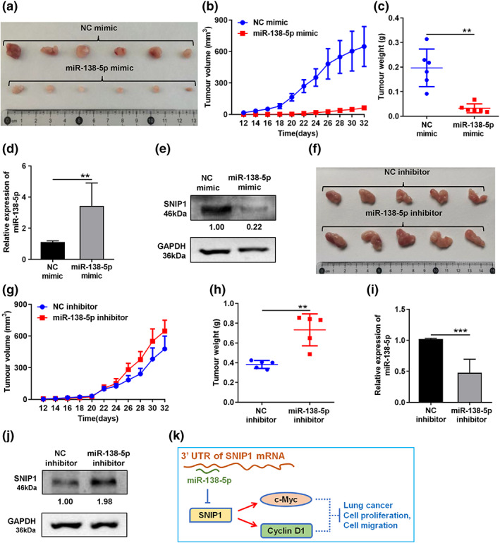
Results: MiR-138-5p was decreased in the serum of patients with non-small cell lung cancer (NSCLC) and in NSCLC cells and tissues. The area under the ROC curve of serum miR-138-5p in the diagnosis of NSCLC was 0.922. This finding indicates the high diagnostic efficiency for lung cancer. MiR-138-5p suppressed but its inhibitor promoted cell proliferation and migration compared with control treatment in vitro and in vivo. MiR-138-5p directly binds to the 3'-untranslated region of SNIP1 and negatively regulated the expression of SNIP1, thereby inhibiting the expression of cyclin D1 and c-Myc. Moreover, overexpression of SNIP1 rescues the miR-138-5p-mediated inhibition in NSCLC cells.
Conclusions: The results suggested that miR-138-5p suppressed lung cancer cell proliferation and migration by targeting SNIP1. Serum miR-138-5p is a novel and valuable biomarker for NSCLC diagnosis.
Keywords: SNIP1; biomarker; lung cancer; miR-138-5p; tumor suppressor.
© 2023 The Authors. Thoracic Cancer published by China Lung Oncology Group and John Wiley & Sons Australia, Ltd.
Conflict of interest statement
The authors declare no conflicts of interest.
Figures

Similar articles
-
The clinical utilization of SNIP1 and its pathophysiological mechanisms in disease.Heliyon. 2024 Jan 17;10(2):e24601. doi: 10.1016/j.heliyon.2024.e24601. eCollection 2024 Jan 30. Heliyon. 2024. PMID: 38304835 Free PMC article. Review.
-
MiR-7-5p suppresses tumor metastasis of non-small cell lung cancer by targeting NOVA2.Cell Mol Biol Lett. 2019 Nov 20;24:60. doi: 10.1186/s11658-019-0188-3. eCollection 2019. Cell Mol Biol Lett. 2019. PMID: 31832068 Free PMC article.
-
UBE2C, Directly Targeted by miR-548e-5p, Increases the Cellular Growth and Invasive Abilities of Cancer Cells Interacting with the EMT Marker Protein Zinc Finger E-box Binding Homeobox 1/2 in NSCLC.Theranostics. 2019 Mar 17;9(7):2036-2055. doi: 10.7150/thno.32738. eCollection 2019. Theranostics. 2019. Retraction in: Theranostics. 2020 Jul 25;10(21):9619. doi: 10.7150/thno.50254. PMID: 31037155 Free PMC article. Retracted.
-
MiR-205-5p promotes lung cancer progression and is valuable for the diagnosis of lung cancer.Thorac Cancer. 2022 Mar;13(6):832-843. doi: 10.1111/1759-7714.14331. Epub 2022 Jan 25. Thorac Cancer. 2022. PMID: 35076182 Free PMC article.
-
Long non-coding RNA PRNCR1 modulates non-small cell lung cancer cell proliferation, apoptosis, migration, invasion, and EMT through PRNCR1/miR-126-5p/MTDH axis.Biosci Rep. 2020 Jul 31;40(7):BSR20193153. doi: 10.1042/BSR20193153. Biosci Rep. 2020. PMID: 31912882 Free PMC article.
Cited by
-
The clinical utilization of SNIP1 and its pathophysiological mechanisms in disease.Heliyon. 2024 Jan 17;10(2):e24601. doi: 10.1016/j.heliyon.2024.e24601. eCollection 2024 Jan 30. Heliyon. 2024. PMID: 38304835 Free PMC article. Review.
-
Extracellular circulating miRNAs as potential non-invasive biomarkers in non-small cell lung cancer patients.Front Oncol. 2023 Jul 21;13:1209299. doi: 10.3389/fonc.2023.1209299. eCollection 2023. Front Oncol. 2023. PMID: 37546401 Free PMC article. Review.
-
Telomere Length, Oxidative Stress Markers, and Related miRNAs in Non-Invasive Samples of Mild COVID-19 Cases.Int J Mol Sci. 2025 May 21;26(10):4934. doi: 10.3390/ijms26104934. Int J Mol Sci. 2025. PMID: 40430074 Free PMC article.
-
Sodium tanshinone IIA sulfonate inhibits tumor growth via miR-138 upregulation in intermittent hypoxia-induced xenograft mice.Aging (Albany NY). 2024 Feb 8;16(4):3231-3240. doi: 10.18632/aging.205531. Epub 2024 Feb 8. Aging (Albany NY). 2024. PMID: 38334965 Free PMC article.
-
PDL1 targeting by miR-138-5p amplifies anti-tumor immunity and Jurkat cells survival in non-small cell lung cancer.Sci Rep. 2024 Jun 12;14(1):13542. doi: 10.1038/s41598-024-62064-5. Sci Rep. 2024. PMID: 38866824 Free PMC article.
References
-
- Sung H, Ferlay J, Siegel RL, Laversanne M, Soerjomataram I, Jemal A, et al. Global cancer statistics 2020: GLOBOCAN estimates of incidence and mortality worldwide for 36 cancers in 185 countries. CA Cancer J Clin. 2021;71(3):209–49. - PubMed
-
- Bernardo BC, Ooi JY, Lin RC, McMullen JR. miRNA therapeutics: a new class of drugs with potential therapeutic applications in the heart. Future Med Chem. 2015;7(13):1771–92. - PubMed
-
- Javadian M, Gharibi T, Shekari N, Abdollahpour‐Alitappeh M, Mohammadi A, Hossieni A, et al. The role of microRNAs regulating the expression of matrix metalloproteinases (MMPs) in breast cancer development, progression, and metastasis. J Cell Physiol. 2019;234(5):5399–412. - PubMed
-
- Baradaran B, Shahbazi R, Khordadmehr M. Dysregulation of key microRNAs in pancreatic cancer development. Biomed Pharmacother. 2019;109:1008–15. - PubMed
Publication types
MeSH terms
Substances
LinkOut - more resources
Full Text Sources
Medical
Molecular Biology Databases
Research Materials
