Video-Assisted Thoracic Surgery Core Needle Biopsy for Pulmonary Nodules in Patients with Impaired Lung Function: Is It Feasible and Safe?
- PMID: 36598118
- PMCID: PMC9845864
- DOI: 10.5090/jcs.22.063
Video-Assisted Thoracic Surgery Core Needle Biopsy for Pulmonary Nodules in Patients with Impaired Lung Function: Is It Feasible and Safe?
Abstract
Background: The number of patients with incidentally identified pulmonary nodules is increasing. This study attempted to confirm the usefulness and safety of video-assisted thoracic surgery (VATS) core needle biopsy of pulmonary nodules.
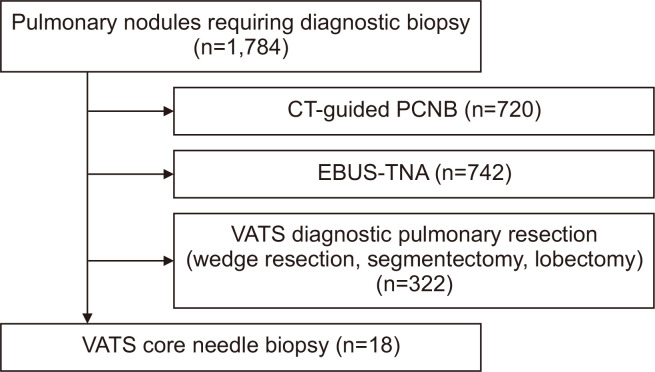
Methods: Data from 18 patients diagnosed with pulmonary nodules who underwent VATS core need biopsy were retrospectively reviewed.
Results: Of the 18 patients, 15 had malignancies (primary lung cancer, n=14; metastatic lung cancer, n=1), and 3 had benign nodules. Mortality and pleural metastasis did not occur during the follow-up period.
Conclusion: In patients with solitary pulmonary nodules that require tissue confirmation, computed tomography-guided percutaneous cutting needle biopsy or diagnostic pulmonary resection sometimes may not be feasible choices due to the location of the solitary pulmonary nodule or the patient's impaired pulmonary function, VATS core needle biopsy may be performed in these patients as an alternative method.
Keywords: Large-core needle biopsy; Solitary pulmonary nodule; Video-assisted thoracic surgery.
Conflict of interest statement
No potential conflict of interest relevant to this article was reported.
Figures
Similar articles
-
Handheld single photon emission computed tomography (handheld SPECT) navigated video-assisted thoracoscopic surgery of computer tomography-guided radioactively marked pulmonary lesions.Interact Cardiovasc Thorac Surg. 2016 Sep;23(3):345-50. doi: 10.1093/icvts/ivw136. Epub 2016 May 20. Interact Cardiovasc Thorac Surg. 2016. PMID: 27207315 Clinical Trial.
-
Video-assisted thoracoscopic solitary pulmonary nodule resection after CT-guided hookwire localization: 43 cases report and literature review.Surg Endosc. 2011 Jun;25(6):1723-9. doi: 10.1007/s00464-010-1502-3. Epub 2010 Dec 22. Surg Endosc. 2011. PMID: 21181200 Review.
-
Needle localization of small pulmonary nodules: Lessons learned.J Thorac Cardiovasc Surg. 2018 May;155(5):2140-2147. doi: 10.1016/j.jtcvs.2018.01.007. Epub 2018 Jan 17. J Thorac Cardiovasc Surg. 2018. PMID: 29455962
-
Computed tomography-guided coil localization for video-assisted thoracoscopic surgery of sub-solid lung nodules: a retrospective study.ANZ J Surg. 2019 Nov;89(11):E514-E518. doi: 10.1111/ans.15450. Epub 2019 Oct 2. ANZ J Surg. 2019. PMID: 31578777
-
[Usefulness of video-assisted thoracoscopy for the diagnosis of solitary pulmonary nodules].Arch Bronconeumol. 2002 Sep;38(9):415-20. doi: 10.1016/s0300-2896(02)75254-x. Arch Bronconeumol. 2002. PMID: 12237012 Review. Spanish.
Cited by
-
Development of a CT image-based virtual atelectasis simulation model and noninvasive lung nodule localization system.J Thorac Dis. 2024 Nov 30;16(11):7651-7662. doi: 10.21037/jtd-24-903. Epub 2024 Nov 21. J Thorac Dis. 2024. PMID: 39678904 Free PMC article.
References
-
- Yang W, Jiang H, Khan AN, et al. Transthoracic needle aspiration in solitary pulmonary nodule. Transl Lung Cancer Res. 2017;6:76–85. doi: 10.21037/tlcr.2017.02.03. https://doi.org/10.21037/tlcr.2017.02.03. - DOI - PMC - PubMed
-
- Nasim F, Ost DE. Management of the solitary pulmonary nodule. Curr Opin Pulm Med. 2019;25:344–53. doi: 10.1097/MCP.0000000000000586. https://doi.org/10.1097/MCP.0000000000000586. - DOI - PubMed
-
- Cruickshank A, Stieler G, Ameer F. Evaluation of the solitary pulmonary nodule. Intern Med J. 2019;49:306–15. doi: 10.1111/imj.14219. https://doi.org/10.1111/imj.14219. - DOI - PubMed
-
- Choo JY, Park CM, Lee NK, Lee SM, Lee HJ, Goo JM. Percutaneous transthoracic needle biopsy of small (≤ 1 cm) lung nodules under C-arm cone-beam CT virtual navigation guidance. Eur Radiol. 2013;23:712–9. doi: 10.1007/s00330-012-2644-6. https://doi.org/10.1007/s00330-012-2644-6. - DOI - PubMed
-
- Verma A, Goh KS, Phua CK, et al. Diagnostic performance of convex probe EBUS-TBNA in patients with mediastinal and coexistent endobronchial or peripheral lesions. Medicine (Baltimore) 2016;95:e5619. doi: 10.1097/MD.0000000000005619. https://doi.org/10.1097/MD.0000000000005619. - DOI - PMC - PubMed
LinkOut - more resources
Full Text Sources