Review
. 2023:279:159-181.
doi: 10.1007/164_2022_624.

CACNA1C-Related Channelopathies

Affiliations
Review

CACNA1C-Related Channelopathies

Kevin G Herold et al. Handb Exp Pharmacol. 2023.

Abstract

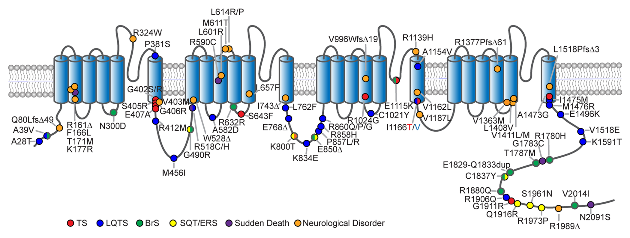
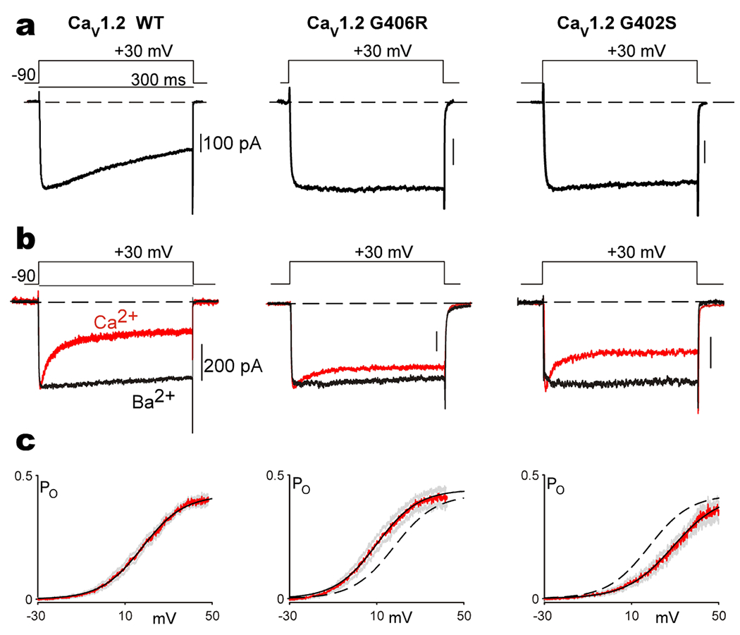
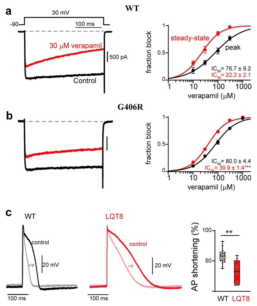
The CACNA1C gene encodes the pore-forming subunit of the CaV1.2 L-type Ca2+ channel, a critical component of membrane physiology in multiple tissues, including the heart, brain, and immune system. As such, mutations altering the function of these channels have the potential to impact a wide array of cellular functions. The first mutations identified within CACNA1C were shown to cause a severe, multisystem disorder known as Timothy syndrome (TS), which is characterized by neurodevelopmental deficits, long-QT syndrome, life-threatening cardiac arrhythmias, craniofacial abnormalities, and immune deficits. Since this initial description, the number and variety of disease-associated mutations identified in CACNA1C have grown tremendously, expanding the range of phenotypes observed in affected patients. CACNA1C channelopathies are now known to encompass multisystem phenotypes as described in TS, as well as more selective phenotypes where patients may exhibit predominantly cardiac or neurological symptoms. Here, we review the impact of genetic mutations on CaV1.2 function and the resultant physiological consequences.

Keywords: CACNA1C; CaV1.2; Channelopathy; L-type calcium channel; Timothy syndrome.


Figures

Figure 1.
Figure 1.. CACNA1C mutations
Cartoon depicting the membrane topology of the pore forming subunit of CaV1.2 indicating the locus of CACNA1C mutations across the channel. Phenotypes associated with each mutation are colored accordingly.
Figure 2.
Figure 2.. TS associated CACANA1C mutations alter the gating of CaV1.2 channels
(a) Exemplar Ba2+ currents measured from CaV1.2 channels display typical VDI, seen as the decay in current over the time course of a 300 ms depolarization. Channels are expressed with the β1B auxiliary subunit, which is permissive of robust VDI. Both the G406R and G402S mutation nearly eliminate VDI in these channels. (b) Exemplar Ba2+ (black) and Ca2+ (red) currents measured from CaV1.2 channels enabling visualization of CDI, seen as the stronger decay in the Ca2+ current as compared to the Ba2+ current. CaV1.2 is expressed with the β2A -auxiliary subunit which decreases the amount of VDI, thus enabling measurement of CDI in relative isolation. Both the G406R and G402S mutations significantly blunt CDI. (c) Open-probability curves measured through single channel recordings of CaV1.2 opening as a function of voltage. Data is displayed in red, with SEM in gray, and a Boltzmann fit to the data shown as the solid black curve. The G406R mutation left shifts the activation of the channel as compared to WT, which is reproduced as the black dashed line (middle panel). The G402S mutation causes a right shift in channel activation (right panel) as compared to the WT data reproduced as the dashed black line. Reproduced with permission from (Dick et. al., 2016, Nat. Commun)(71).
Figure 3.
Figure 3.. CACANA1C mutations reduce the efficacy of verapamil
(a) Verapamil concentration-response curves measured for WT CaV1.2 in HEK 293 cells. Left: Exemplar Ba2+ current before (black) and after (red) the addition of 30 μM verapamil. Accumulation of block can be seen during the 300 ms depolarization in the presence of verapamil (red). Right: concentration-response curves can be generated by measuring the effect of the drug on the peak current amplitude immediately following depolarization (black), or at the end of the voltage step after 300 ms depolarization (red). The increased block measured from the steady-state value (red) is indicative of use-dependence of the drug. (b) The G406R mutation significantly reduces the use-dependent block of verapamil, seen as an increase in steady-state IC50. (c) Left: Exemplar AP recordings of WT iPSC derived cardiomyocytes before (black) and after (gray) the addition of 10 μM verapamil. Middle: Exemplar AP recording from an iPSC derived cardiomyocyte from a patient harboring the G406R mutation (red, LQT8) shows significant prolongation of the action potential as compared to WT (left, black). Addition of 10 μM verapamil (pink) reduces the AP duration to a lesser extent as compared to WT iPSC derived cardiomyocytes. Right: Quantification of the percent of AP shortening in WT vs. LQT8 (G406R) containing iPSC derived cardiomyocytes demonstrates a significant deficit in verapamil’s ability to shorten the AP in the context of patient derived cells harboring the G406R mutation (LQT8). Reproduced with permission from (Dick et. al., 2022, JMCC)(137).

References

    1. Berger SM, Bartsch D, The role of L-type voltage-gated calcium channels Cav1.2 and Cav1.3 in normal and pathological brain function. Cell Tissue Res 357, 463–476 (2014). - PubMed
    1. Harvey RD, Hell JW, CaV1.2 signaling complexes in the heart. J Mol Cell Cardiol 58, 143–152 (2013). - PMC - PubMed
    1. Zamponi GW, Striessnig J, Koschak A, Dolphin AC, The Physiology, Pathology, and Pharmacology of Voltage-Gated Calcium Channels and Their Future Therapeutic Potential. Pharmacol Rev 67, 821–870 (2015). - PMC - PubMed
    1. Bers DM, Ca regulation in cardiac muscle. Med Sci Sports Exerc 23, 1157–1162 (1991). - PubMed
    1. Lederer WJ et al., Excitation-contraction coupling in heart cells. Roles of the sodium-calcium exchange, the calcium current, and the sarcoplasmic reticulum. Annals of the New York Academy of Sciences 588, 190–206 (1990). - PubMed

Substances

Supplementary concepts