Association between serum periostin levels and the severity of arsenic-induced skin lesions
- PMID: 36598904
- PMCID: PMC9812306
- DOI: 10.1371/journal.pone.0279893
Association between serum periostin levels and the severity of arsenic-induced skin lesions
Abstract
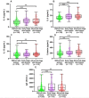
Arsenic is a potent environmental toxicant and human carcinogen. Skin lesions are the most common manifestations of chronic exposure to arsenic. Advanced-stage skin lesions, particularly hyperkeratosis have been recognized as precancerous diseases. However, the underlying mechanism of arsenic-induced skin lesions remains unknown. Periostin, a matricellular protein, is implicated in the pathogenesis of many forms of skin lesions. The objective of this study was to examine whether periostin is associated with arsenic-induced skin lesions. A total of 442 individuals from low- (n = 123) and high-arsenic exposure areas (n = 319) in rural Bangladesh were evaluated for the presence of arsenic-induced skin lesions (Yes/No). Participants with skin lesions were further categorized into two groups: early-stage skin lesions (melanosis and keratosis) and advanced-stage skin lesions (hyperkeratosis). Drinking water, hair, and nail arsenic concentrations were considered as the participants' exposure levels. The higher levels of arsenic and serum periostin were significantly associated with skin lesions. Causal mediation analysis revealed the significant effect of arsenic on skin lesions through the mediator, periostin, suggesting that periostin contributes to the development of skin lesions. When skin lesion was used as a three-category outcome (none, early-stage, and advanced-stage skin lesions), higher serum periostin levels were significantly associated with both early-stage and advanced-stage skin lesions. Median (IQR) periostin levels were progressively increased with the increasing severity of skin lesions. Furthermore, there were general trends in increasing serum type 2 cytokines (IL-4, IL-5, IL-13, and eotaxin) and immunoglobulin E (IgE) levels with the progression of the disease. The median (IQR) of IL-4, IL-5, IL-13, eotaxin, and IgE levels were significantly higher in the early-and advanced-stage skin lesions compared to the group of participants without skin lesions. The results of this study suggest that periostin is implicated in the pathogenesis and progression of arsenic-induced skin lesions through the dysregulation of type 2 immune response.
Copyright: © 2023 Khatun et al. This is an open access article distributed under the terms of the Creative Commons Attribution License, which permits unrestricted use, distribution, and reproduction in any medium, provided the original author and source are credited.
Conflict of interest statement
The authors have declared that no competing interests exist.
Figures


References
-
- Mukherjee SC, Saha KC, Pati S, Dutta RN, Rahman MM, Sengupta MK, et al.. Murshidabad—one of the nine groundwater arsenic-affected districts of West Bengal, India. Part II: dermatological, neurological, and obstetric findings. Clin Toxicol (Phila). 2005; 43:835–848. doi: 10.1080/15563650500357495 - DOI - PubMed
-
- Chakraborti D, Rahman MM, Das B, Chatterjee A, Das D, Nayak B, et al.. Groundwater arsenic contamination and its health effects in India. Hydrogeol J. 2017; 25:1165–1181. 10.1007/s10040-017-1556-6 - DOI
Publication types
MeSH terms
Substances
Associated data
Grants and funding
LinkOut - more resources
Full Text Sources
Medical

