. 2023 Jan 10;120(2):e2213418120.
doi: 10.1073/pnas.2213418120. Epub 2023 Jan 4.

Re-formation of synaptic connectivity in dissociated human stem cell-derived retinal organoid cultures

Affiliations

Re-formation of synaptic connectivity in dissociated human stem cell-derived retinal organoid cultures

Allison L Ludwig et al. Proc Natl Acad Sci U S A. .

Abstract

Human pluripotent stem cell (hPSC)-derived retinal organoids (ROs) can efficiently and reproducibly generate retinal neurons that have potential for use in cell replacement strategies [Capowski et al., Development 146, dev171686 (2019)]. The ability of these lab-grown retinal neurons to form new synaptic connections after dissociation from ROs is key to building confidence in their capacity to restore visual function. However, direct evidence of reestablishment of retinal neuron connectivity via synaptic tracing has not been reported to date. The present study employs an in vitro, rabies virus-based, monosynaptic retrograde tracing assay [Wickersham et al., Neuron 53, 639-647 (2007); Sun et al., Mol. Neurodegener. 14, 8 (2019)] to identify de novo synaptic connections among early retinal cell types following RO dissociation. A reproducible, high-throughput approach for labeling and quantifying traced retinal cell types was developed. Photoreceptors and retinal ganglion cells-the primary neurons of interest for retinal cell replacement-were the two major contributing populations among the traced presynaptic cells. This system provides a platform for assessing synaptic connections in cultured retinal neurons and sets the stage for future cell replacement studies aimed at characterizing or enhancing synaptogenesis. Used in this manner, in vitro synaptic tracing is envisioned to complement traditional preclinical animal model testing, which is limited by evolutionary incompatibilities in synaptic machinery inherent to human xenografts.

Keywords: human pluripotent stem cells; photoreceptors; retinal organoid; synapses; trans-synaptic tracing.


Conflict of interest statement

The authors declare competing interests and have patent filings to disclose. D.M.G. has declared intellectual rights for production of 3D ROs through the Wisconsin Alumni Research Foundation, Madison, WI (US PTO No. 9,328,328). The authors have organizational affiliations and research support to disclose. D.M.G. has an ownership interest in and receives grant support from Opsis Therapeutics LLC, which has licensed the technology to generate ROs from hPSCs utilized in this publication. The terms of this arrangement have been reviewed and approved by the University of Wisconsin-Madison in accordance with its conflict-of-interest policies. All other authors declare no competing interests.

Figures
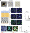
Fig. 1.
Fig. 1.
Retinal neuron diversity in dissociated hPSC-derived RO cultures. (A) Experimental overview showing the appearance of (Left) an intact stage 2 RO (D89 shown), (Middle) retinal cells immediately following dissociation of a D80 RO, and (Right) dissociated retinal cells 20 d after plating. (B) ICC showing the relative appearance and qualitative abundance of SNCG+ (RGCs), CALB+ (retinal interneurons), and RCVN+ (maturing photoreceptors) cells in dissociated D80 RO cultures 20 d after plating, alongside a schematic depicting their location in intact retinal tissue. (C) Quantification of retinal cell populations expressing SNCG, CALB, RCVN, Ki67 (dividing cells, including retinal progenitors and RPE cells), and/or CRX (newborn and maturing photoreceptors) in dissociated D80 RO cultures 20 d after plating. (D) RCVN+ photoreceptors express the synaptic proteins VGLUT1 and SYNAP 20 d after D80 RO dissociation and plating, which colocalize with each other along photoreceptor axons (arrows). Scale bars: 100 µm, 50 µm, or 10 µm as indicated on their respective panels. (E) Quantification of RCVN+ cells possessing processes with VGLUT1+ puncta at day 1, 2, 5, and 20 d postplating. At day 1, RCVN+ cells (n = 467) displayed rare processes, none of which contained VGLUT1+ puncta. Thereafter, the percentage of RCVN+ cells having processes with VGLUT1+ puncta increased substantially (day 2: 1.4 ± 0.6%, n = 559 cells; day 5: 8.4 ± 1.4%, n = 633 cells; day 20: 29.5 ± 7.1%, n = 749 cells) (*P < 0.05, Kruskal–Wallis with pairwise Mann–Whitney U analyses).
Fig. 2.
Fig. 2.
Monosynaptic retrograde rabies virus tracing assay design and validation. (A) Schematic depicting the two-step RaV synaptic tracing assay utilized for this study, alongside a fluorescence image from one of the experiments (Far Right) showing an example of a starter retinal cell (arrow) and a traced presynaptic retinal cell (arrowhead). ROs were dissociated and plated on day 80 of differentiation. Resulting 2D cultures were infected with lentivirus 10 d later (day 90), followed by RaV infection at day 95 and examination for the presence of starter and traced presynaptic cells at day 100. (B) In the absence of primary transduction with lenti-GTR (i.e., no lentivirus control), secondary RaV infection cannot occur, resulting in negligibly detectable fluorescent cells. (C) Primary transduction with lenti-GTΔR followed by secondary RaV infection permits labeling of starter retinal cells (GFP+ nuclei and mCherry+ cytoplasm) (arrows) but does not allow true monosynaptic tracing. Therefore, the small fraction of cells that have GFP-negative nuclei and mCherry+ cytoplasm in these control experiments represent nonspecific biomaterial transfer (i.e., false traced presynaptic cell detection rate). (D) In experimental cultures, primary transduction with lenti-GTR followed by secondary RaV infection leads to labeling of both starter retinal cells and traced presynaptic retinal cells (arrows). The median percentage of traced presynaptic cells was significantly greater in lenti-GTR + RaV cultures [6.2% (95% CI: 5.6, 6.9) of DAPI+ cells, n = 45 replicate wells] (D) relative to lenti-GTΔR + RaV controls [1.4% (95% CI: 1.1, 1.9) of DAPI+ cells, n = 37 replicate wells] (C) or RaV-only controls [0.02% (95% CI: 0.01, 0.04) of DAPI+ cells, n = 34 replicate wells]; P < 0.00001 using Mood’s test of equal medians. Scale bars: 100 µm, 50 µm as indicated on respective panels.
Fig. 3.
Fig. 3.
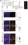
Monosynaptic retrograde rabies virus tracing reveals new synaptic contacts formed by photoreceptors, interneurons, and retinal ganglion cells after dissociation from ROs. (A) ICC images showing representative traced presynaptic cells (arrows) alongside a schematic depicting their corresponding location within intact retinal tissue (Top panels: SNCG+ retinal ganglion cells; Middle panels: CALB+ retinal interneuron; Lower panels: RCVN+ photoreceptor). (B) Quantification of traced presynaptic cell populations coexpressing markers of proliferation (Ki67), retinal ganglion cells (SNCG), retinal interneurons (CALB), or photoreceptors (CRX, RCVN). Error bars represent SDs. Scale bars: 100 µm or 50 µm as indicated on respective panels.

Similar articles

Cited by

References

    1. Cajal S. R. y., Estructura de los centros nerviosos de las aves. Revista Trimestral de Histología Normal y Patológica, 1, 1–10 (1888).
    1. Soto F., Kerschensteiner D., Synaptic remodeling of neuronal circuits in early retinal degeneration. Front. Cell. Neurosci. 9, 395 (2015). - PMC - PubMed
    1. Jones B. W., Marc R. E., Retinal remodeling during retinal degeneration. Exp. Eye Res. 81, 123–137 (2005). - PubMed
    1. Quigley H. A., Glaucoma. Lancet 377, 1367–1377 (2011). - PubMed
    1. Ribeiro J., et al. , Restoration of visual function in advanced disease after transplantation of purified human pluripotent stem cell-derived cone photoreceptors. Cell Rep. 35, 109022 (2021). - PMC - PubMed

Publication types

Substances