. 2023 Jan 4;6(3):e202201594.
doi: 10.26508/lsa.202201594. Print 2023 Mar.

An essential Noc3p dimerization cycle mediates ORC double-hexamer formation in replication licensing

Affiliations

An essential Noc3p dimerization cycle mediates ORC double-hexamer formation in replication licensing

Aftab Amin et al. Life Sci Alliance. .

Abstract

Replication licensing, a prerequisite of DNA replication, helps to ensure once-per-cell-cycle genome duplication. Some DNA replication-initiation proteins are sequentially loaded onto replication origins to form pre-replicative complexes (pre-RCs). ORC and Noc3p bind replication origins throughout the cell cycle, providing a platform for pre-RC assembly. We previously reported that cell cycle-dependent ORC dimerization is essential for the chromatin loading of the symmetric MCM double-hexamers. Here, we used Saccharomyces cerevisiae separation-of-function NOC3 mutants to confirm the separable roles of Noc3p in DNA replication and ribosome biogenesis. We also show that an essential and cell cycle-dependent Noc3p dimerization cycle regulates the ORC dimerization cycle. Noc3p dimerizes at the M-to-G1 transition and de-dimerizes in S-phase. The Noc3p dimerization cycle coupled with the ORC dimerization cycle enables replication licensing, protects nascent sister replication origins after replication initiation, and prevents re-replication. This study has revealed a new mechanism of replication licensing and elucidated the molecular mechanism of Noc3p as a mediator of ORC dimerization in pre-RC formation.


Conflict of interest statement

The authors declare that they have no conflict of interest.

Figures
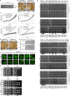
Figure S1.
Figure S1.. Additional data together with Fig 1 to verify the NOC3 separation-of-function (replication versus ribosome biogenesis) mutants; Noc3p is essential for cell growth and survival; NOC3 and noc3-1-ribo, but not noc3-3-rep, are multicopy suppressors of the mcm5-1 and orc5-1 mutant cells.
(A) Noc3p was mostly degraded by 4 h and was no longer detectable by 6 h after the GAL-NOC3 cells were shifted from galactose-containing medium (YPG) to glucose-containing medium (YPD). Mcm2p and Orc3p were used for comparison. (B) W303-1A, GAL-NOC3, GAL-NOC3/pRS416, and GAL-NOC3/pRS416-NOC3 cells were streaked on YPD and YPG plates. The plates were incubated at 25°C for 3–5 d. (C) Growth curves of the WT and NOC3 ts mutants when the asynchronous cells were cultured at 25°C or shifted to 37°C. Cell densities were measured at one-hour intervals. Results were the average ± SD of three independent experiments. (D) W303-1A, noc3-3-rep, noc3-1-ribo, and noc3-ts-URA-both cells were streaked onto YPD plates and incubated at 25°C or 37°C for 3–5 d. (E) 10-fold serial dilutions of noc3-3-rep cells containing pRS416 or pRS416-NOC3 plasmid were spotted onto SCM-Ura (synthetic complete medium lacking uracil) plates and grown at 25°C or 37°C for 3–5 d. (F) Microscopic images of nuclear-accumulated Rpl25-GFP in G1-phase–synchronized WT cells and those of five different NOC3 mutant strains grown at 25°C and 37°C (n ≥ 300 for each cell sample). Localization was visualized under a fluorescence microscope within 10 min of harvesting. Scale bar, 10 µm. Magnification, 600×. Arrowheads indicate nuclear accumulation. The NOC3 mutants are denoted with a postfix “-rep” (severely defective in DNA replication with mild defects in ribosome biogenesis), “-ribo” (severely defective in ribosome biogenesis without obvious defect in DNA replication), or “-both” (with intermediate levels of deficiencies in both ribosome biogenesis and DNA replication), for easy reference. (G) 10-fold serial dilutions of NOC3, noc3-1-ribo, noc3-3-rep, noc3-9-rep, and noc3-142-both cells containing the plasmids overexpressing the indicated initiation proteins were spotted onto plates containing galactose or glucose and grown at 25°C. The open diamonds indicate that the genes induced dominant-negative effects in all strains. The closed diamonds indicate that the genes caused NOC3 mutant allele-specific dosage lethality phenotypes. The closed circles indicate that the genes induced dominant-negative effects in all replication-deficient NOC3 mutant strains. (H) 10-fold serial dilutions of the mcm5-1 and orc5-1 mutant cells transformed with the multicopy plasmids pRS425-NOC3, pRS425-noc3-1, or pRS425-noc3-3, or the vector pRS425, were spotted on SCM-Leu (synthetic complete medium lacking leucine) plates and grown at 25°C or 37°C for 4 d. Untransformed strains (upper panels) were used as controls to show the similar temperature sensitivities of the mutants.
Figure 1.
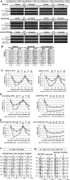
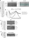
Figure 1.. NOC3 separation-of-function mutants show differential defects in DNA replication initiation and ribosome biogenesis.
(A) Ribosome profiles (OD260) for the WT and five NOC3 ts strains synchronized in G1-phase by α-factor before being shifted to 37°C. Equal amounts of cell lysates were fractionated through 20–60% sucrose gradients. The 40S small subunit, 60S large subunit, and 80S mono-ribosome and poly-ribosome were indicated by arrows. A normal polysome profile is indicated as 100%, whereas other values show different levels of polysome defects. The OD260 reading of sucrose gradient fractions was measured using a spectrophotometer. The averages from three independent experiments are shown. (B) Quantification of the nuclear-accumulated Rpl25-GFP in WT and six different NOC3 mutants at 25°C and 37°C (n ≥ 300 for each cell sample). (C) Quantitative plasmid loss rates were measured for the WT and six NOC3 strains containing either p1ARS or p8ARSs grown in YPRG medium at 30°C as indicated. Plasmid loss rates are expressed as the average percentage of loss per generation. Results were the average ± SD of three independent experiments. Statistical analysis was carried out by one-Way ANOVA and Dunnett’s multiple comparison test. Not significant (n/s), P > 0.05; *, P < 0.05; **, P < 0.01; and ***, P < 0.001. The NOC3 mutants are denoted with a postfix “-rep” (severely defective in DNA replication with mild defects in ribosome biogenesis), “-ribo” (severely defective in ribosome biogenesis without obvious defect in DNA replication), or “-both” (with intermediate levels of deficiencies in both ribosome biogenesis and DNA replication), for easy reference.
Figure S2.
Figure S2.. Additional data together with Fig 2 to show that Noc3p is required for ORC dimerization, MCM loading, and cell cycle progression; G1/S transition is impeded in noc3-3-rep cells at the non-permissive temperature.
(A) Cells of the indicated strains were pre-synchronized in G1-phase with α-factor (α-F./αF), released into fresh medium, and re-synchronized in nocodazole (Noc./0′) at 25°C and 37°C. Cells were released into fresh medium at 25°C or 37°C and harvested at the indicated time points for chromatin-binding assays to detect the chromatin-bound Mcm2p, Orc3p, and histone H3 (for chromatin)/β-actin (for soluble proteins). *, anti-Orc3 cross-reacting band. (A, B) Corresponding flow cytometry analysis for the chromatin-binding assay experiments shown in (A). Asy., asynchronous cells. (A, C, D, E) Quantification of the chromatin levels of Orc3p and Mcm2p for the experiments shown in (A) for W303-1A (C), noc3-9-rep (D), and noc3-ts-URA-both (E) cells, presented as the average ± SD of three independent experiments. The signals of Orc3p and Mcm2p were normalized to that of histone H3 at different time points, and the resulting numbers were then further normalized to the G1-phase sample (αF). Statistical analysis was carried out by a paired t test, comparing data with those of 0′ time point (Noc.). *, Orc3p; #, Mcm2p; *n/s and #n/s, not significant with P > 0.05; */#, P < 0.05; **/##, P < 0.01; and ***/###, P < 0.001. (F) α-Factor–synchronized cells of the indicated strains at 25°C were released into fresh medium at 25°C, and cell samples were collected at 30-min intervals. (G) α-Factor–synchronized cells of the indicated strains at 25°C were shifted to 37°C for 3 h and then released into fresh medium at 37°C. Cell samples were collected at 30-min intervals.
Figure 2.
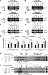
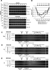
Figure 2.. NOC3 separation-of-function (replication versus ribosome biogenesis) mutants; Noc3p is required for ORC dimerization, MCM loading, and cell cycle progression.
(A) Indicated mutant cells were pre-synchronized in G1-phase with α-factor (α-F.), released into fresh medium, and re-synchronized in nocodazole (Noc./0′) at the permissive (25°C) and non-permissive temperatures (37°C). Cells were released into the cell cycle and harvested at the indicated time points and temperatures for chromatin-binding assay to detect the chromatin-bound levels of Mcm2p, Orc3p, and histone H3 (for the chromatin factions)/β-actin (for the soluble proteins). (A, B) Corresponding flow cytometry for the chromatin-binding assay experiments shown in (A). (A, C, D) Quantification of the chromatin levels of Orc3p and Mcm2p for the 25°C and 37°C experiments shown in (A) presented as the average ± SD of three independent experiments. The signals of Orc3p and Mcm2p were normalized to that of histone H3 at different time points, and the resulting numbers were then further normalized to the G1-phase sample (αF). Statistical analysis was carried out by a paired t test (signals of time points versus those in nocodazole). *, Orc3p; #, Mcm2p; not significant (*n/s, #n/s), P > 0.05; */#, P < 0.05; **/# #, P < 0.01; and ***/###, P < 0.001.
Figure 3.
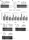
Figure 3.. Noc3p is required for ORC dimerization in G1-phase, and large molecular forms of endogenous ORC do not exist in G1-synchronized noc3-3-rep cells at 37°C.
(A, B, C, D, E, F) Extracts from G1-phase–synchronized noc3-1-ribo (A, B), noc3-3-rep (C, D), and noc3-ts-URA-both (E, F) mutant cells expressing Myc-Orc2 and Orc2-FLAG (A, C, E), or Myc-Orc6 and Orc6-FLAG (B, D, F), grown at 25°C or shifted to 37°C, were immunoprecipitated with anti-FLAG antibody or control mouse IgG. Whole-cell extracts (input) and immunoprecipitates by α-FLAG or control IgG were immunoblotted with anti-FLAG and anti-Myc antibodies. (G) Sequential ChIP (re-ChIP) assays were performed with G1-synchronized noc3-1-ribo or noc3-3-rep cells co-expressing Orc6-FLAG and Myc-Orc6 shifted to 37°C. Extracts were first immunoprecipitated with anti-FLAG antibody. The anti-FLAG ChIP chromatin immunoprecipitates were eluted and then immunoprecipitated with anti-Myc antibody in the second ChIP. Real-time PCR was performed using primers to quantify ARS1, ARS1 + 2.5 kb, ARS501, ARS501 + 11 kb, ARS305, and ARS305 + 8 kb. The relative enrichment (ARS normalized to non-ARSs) was calculated and averaged from three independent experiments. Data are presented as the mean ± SD. Statistical analysis was carried out by one-Way ANOVA and Dunnett’s multiple comparison test. Not significant (n/s), P > 0.05; *, P < 0.05; **, P < 0.01; and ***, P < 0.001. (H, I) noc3-3-rep mutant cells synchronized in G1-phase at 25°C (H) and those shifted to 37°C (I) were cross-linked, harvested, and used to prepare total protein extracts for 20–60% sucrose gradient analysis. Alkaline phosphatase (150 kD) and β-galactosidase (450 kD) were applied as protein markers. The resulting 26 fractions and the gradient load from each cell sample were resolved by 10% SDS–PAGE and were immunoblotted with anti-Orc3 and anti-Mcm2 antibodies. Flow cytometry was used to determine the cell cycle distribution of the cells. The immunoblots are representative images from one of the three independent experiments that produced similar results.
Figure S3.
Figure S3.. Additional data together with Fig 3 to show that Noc3p is required for the formation of larger molecular forms of endogenous ORC in asynchronous and G1-phase cells but not M-phase cells
(A, B, C, D, E, F, G, H, I, J) Mutant noc3-1-ribo cells growing asynchronously or synchronized (G1-phase/M-phase) at 25°C (A, B, C) and those shifted to 37°C (D, E, F), and noc3-3-rep mutant cells grown asynchronously or synchronized in M-phase at 25°C (G, H) and those shifted to 37°C (I, J) were cross-linked with formaldehyde, harvested, and used to prepare total protein extracts for 20–60% sucrose gradient analysis. Alkaline phosphatase (150 kD) and β-galactosidase (450 kD) were applied as protein markers. The resulting 26 fractions and the gradient load from each cell sample were run on 10% SDS–PAGE and were immunoblotted with anti-Orc3 and anti-Mcm2 antibodies. Flow cytometry was used to determine the cell cycle distribution of the cells. The immunoblots are representative images from three independent experiments that produced similar results.
Figure 4.
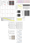
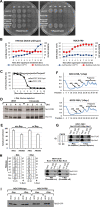
Figure 4.. Noc3p self-interacts and dimerizes in a cell cycle–regulated manner, and deletion of the Noc3p self-interaction domain impairs self-interaction but not interaction with ORC.
(A) Extracts from cycling cells expressing Myc-Noc3 and Noc3-FLAG were immunoprecipitated with anti-FLAG antibody, anti-Myc antibody, or control mouse IgG. Whole-cell extracts (input) and immunoprecipitates (IP) were immunoblotted with anti-FLAG and anti-Myc antibodies. Note that the Myc-Noc3p signal in lane 1 is slightly distorted. (B) Extracts from cells expressing Noc3-FLAG and Noc3-Myc in G1-, S-, or M-phase were immunoprecipitated by anti-FLAG and probed for Noc3-FLAG and Noc3-Myc. Flow cytometry data for the co-IP are also shown. (C) Sequential ChIP (re-ChIP) assays were performed with G1-, S-, or M-phase–synchronized cells co-expressing Noc3-FLAG and Myc-Noc3. Extracts were first immunoprecipitated with anti-FLAG antibody. The anti-FLAG chromatin immunoprecipitates were then eluted and immunoprecipitated with anti-Myc antibody in the second ChIP. Real-time PCR was performed using primers to quantify ARS1, ARS1 + 2.5 kb, ARS501, ARS501 + 11 kb, ARS305, and ARS305 + 8 kb. The relative enrichment (ARS normalized to non-ARSs) was calculated and averaged from three independent experiments. Data are presented as the mean ± SD. Statistical analysis was carried out by one-Way ANOVA and Dunnett’s multiple comparison test. Not significant (n/s), P > 0.05; *, P < 0.05; **, P < 0.01; and ***, P < 0.001. (D, E, F) Extracts from asynchronous cells expressing Noc3-CC2∆-FLAG and Myc-Noc3-CC2∆ (D), and those expressing Noc3-CC2∆-FLAG only (E, F), were immunoprecipitated with the indicated antibodies or control mouse IgG. Whole-cell extracts (input) and immunoprecipitates (Co-IP) were immunoblotted with the indicated antibodies.
Figure S4.
Figure S4.. Additional data together with Fig 4 to show that the CC2 domain of Noc3p is conserved across species; CC2 is required for Noc3p self-interaction but not for interaction with ORC; CC2 is essential for S-phase entry but not DNA replication elongation; and ribosome biogenesis is not affected by the Noc3p truncation or the NOC3 endogenous gene shutoff within the experimental time window.
(A) Schematic diagram of the NOC3 fragments studied and mapping of the Noc3p self-interaction domains by the yeast two-hybrid assay. Interactions between pairs of BD- and AD-fusion proteins were examined by yeast two-hybrid analysis on SCM-Trp-Leu (non-selective for the protein–protein interactions examined) and SCM-Trp-Leu-His (selective for the protein–protein interactions examined) plates. As negative controls, the host yeast cells were co-transformed with the empty BD or AD vector together with the individual fusion proteins as indicated. (B) Results of MultiCoil analysis of NOC3 from different species. Arrowheads indicate the conserved C-terminal coiled-coil domains (CC2). (C) Similarity comparison of NOC3 across different species. (D) Multiple alignments of the C-terminal coiled-coil region of NOC3 from different species: H. sapiens (Homo), P. troglodytes (Pan), P. pygmaeus (Pongo), M. musculus (Mus), R. norvegicus (Rattus), G. gallus (Gallus), D. rerio (Danio), D. melanogaster (Drosophila), and S. cerevisiae (Saccharomyces). (E) Schematic diagram of the Noc3p fragments studied. A summary table lists the relative strengths of the respective interactions observed by the host yeast cell growth on SCM-Trp-Leu-His plates. (F) 10-fold serial dilutions of GAL-NOC3 cells harboring the empty vector or the plasmid expressing WT Noc3p, noc3-CC1Δ, or noc3-CC2Δ. The cells were spotted onto galactose- or glucose-containing plates. (G) Yeast two-hybrid assays for the interactions of BD-NOC3 and BD-Noc3-CC2Δ with AD-Noc3p, AD-Orc2p, and AD-Orc3p. (H) Schematic diagram of the NOC3 domain deletion mutants studied, the resulting growth phenotypes of the cells expressing the WT or Noc3p deletion mutants, and the relative strengths of the respective interactions of the WT or mutant proteins observed by the host yeast cell growth on SCM-Trp-Leu-His plates. (I) Expression of the endogenous Noc3p in GAL-NOC3 cells, containing the empty vector, WT NOC3, noc3-CC1Δ, or noc3-CC2Δ plasmids, was shut down in glucose-containing medium for 4 h before the cells were synchronized in G1-phase by α-factor (α-F.). Cells were then released into fresh glucose-containing medium. Flow cytometry was performed with the cell samples taken at the indicated time points. Asy., asynchronous cells. (J) GAL-NOC3 cells containing the empty vector or the plasmid expressing Noc3p, Noc3-CC1Δ, or Noc3-CC2Δ were grown in glucose-containing medium for 1.5 h (to suppress the Noc3p expression from GAL-NOC3) before being synchronized in G1-phase with α-factor. The cells were then released from the G1-phase block into early S-phase in hydroxyurea (HU)-containing medium with glucose for 2.5 h. Afterward, the cells were released into fresh medium containing glucose. Flow cytometry was performed with the cell samples taken at the indicated time points. (K) Quantification of the nuclear-accumulated Rpl25-GFP in the indicated strains. (I) GAL-NOC3 cells containing the indicated plasmids were cultured as described in (I), and the Rpl25-GFP localization was examined before and after the cells were shifted to glucose-containing medium. (F, G, K, L) Ribosome profiles (OD260) for the strains described in (F, G, K) after the cells were shifted to glucose-containing medium. Equal amounts of cell lysates were fractionated through on 20–60% sucrose gradients. The 40S small subunit, 60S large subunit, and 80S mono-ribosome and poly-ribosome were indicated by arrows. (M) NOC domain deletion ts mutant cells containing the empty vector or the plasmid expressing the WT Noc3p, Noc3-NOCΔ mutant, or noc3-CC2Δ mutant proteins were grown on galactose- or glucose-containing plates at the indicated temperatures.
Figure 5.
Figure 5.. Free Noc3p loads onto chromatin to form double-hexamers in late M-phase before MCM loading.
(A) NOC3-HA cells were pre-synchronized in G1-phase with α-factor (α-F./αF) and then released into fresh medium. The cells were then re-synchronized in M-phase with nocodazole (Noc./0′). The cells were subsequently released into the cell cycle for chromatin-binding assay to detect the chromatin-bound Mcm2p, Noc3p-HA, Orc3p, and histone H3 (for chromatin) or β-actin (for soluble proteins). *, anti-Orc3 cross-reacting band. (A, B) Quantification of the chromatin levels of Mcm2p, Noc3p-HA, and Orc3p for the experiments shown in (A), presented as the average ± SD of three independent experiments. The signals of Orc3p, Noc3p-HA, and Mcm2p were normalized to that of histone H3 at different time points, and the resulting numbers were then further normalized to the G1-phase sample (αF). Statistical analysis was carried out by a paired t test, comparing data with those of the 0′ time point (Noc.). (^, Noc3p-HA; *, Orc3p; #, Mcm2p; not significant [^n/s, *n/s, #n/s], P > 0.05; ^/*/#, P < 0.05; ^^/**/##, P < 0.01; and ^^^/***/###, P < 0.001). (C) GAL-Myc-NOC3 cells were synchronized in G1-phase, induced to express Myc-Noc3, and then released into the cell cycle. Samples were collected at the indicated time points. Whole-cell extracts and DNase I–solubilized chromatin fractions were immunoblotted for Mcm2p, Myc-Noc3, and histone H3. (D) W303-1A (WT) cells expressing Noc3-FLAG and Myc-Noc3 were synchronized in M-phase and released into G1-phase in fresh medium containing α-factor. Cell samples were harvested at the indicated time points for co-IP. The cell cycle stages as marked were determined by flow cytometry.
Figure S5.
Figure S5.. Additional data together with Fig 5 to show that Noc3p self-interacts and dimerizes in a cell cycle–regulated manner, and the existence of larger molecular forms of endogenous Noc3p in asynchronous and G1-phase cells, but not M-phase cells.
(A) NOC3-HA cells were arrested in G1-phase with α-factor and then released into fresh medium. Cells from equal culture volumes were harvested at the indicated time points for chromatin-binding assay to detect the chromatin-bound Mcm2p, Noc3p-HA, Orc3p, and histone H3 (for chromatin) or β-actin (for supernatant). *, anti-Orc3 cross-reacting band. (A, B) Quantification of the chromatin levels of Orc3p, Mcm2p, and Noc3-HA for the experiments shown in (A), presented as the average ± SD of three independent experiments. The signals of Orc3p, Noc3p-HA, and Mcm2p were normalized to that of histone H3 at different time points, and the resulting numbers were then further normalized to the G1-phase sample (αF). Statistical analysis was carried out by a paired t test (each time point versus that in α-factor block). ^, Noc3-HA; *, Orc3p; #, Mcm2p; not significant (^n/s, *n/s, #n/s), P > 0.05; ^, *, #, P < 0.05; ^^, **, # #, P < 0.01; and ^^^, ***, ###, P < 0.001. (C, D, E) NOC3-HA cells growing asynchronously or synchronized (G1-phase or M-phase) were cross-linked, harvested, and used to prepare total protein extracts for 20–60% sucrose gradient analysis. Alkaline phosphatase (150 kD) and β-galactosidase (450 kD) were applied as protein markers. The resulting 26 fractions and the gradient load from each cell sample were run on 10% SDS–PAGE and were immunoblotted with anti-HA, anti-Orc3, and anti-Mcm2 antibodies. Flow cytometry was used to determine the cell cycle distribution of the cells. The immunoblots are representative images from three independent experiments that produced similar results.
Figure S6.
Figure S6.. Additional data together with Fig 6 to show that depletion of non–chromatin-bound Noc3p from the nucleus during the M-to-G1 transition impairs Noc3p self-interaction, DNA replication, cell proliferation, and cell viability; the anchor-away system does not impair chromatin-bound Noc3p or ribosome biogenesis; the anchor-away system does not cause steric hindrance on chromatin from the ribosome; and Noc3p dimerization in G1-phase is independent of ORC dimerization and Cdc6p.
(A) 10-fold serial dilutions of cells from HHY212 (NOC3 WT) and NOC3-FRB strains. The cells, containing the empty vector or a plasmid expressing the WT NOC3, were spotted on galactose-containing plates with or without rapamycin and grown at 30°C for 3 d. (B) HHY212 (NOC3 WT) and NOC3-FRB cells were treated with rapamycin. Cell number (circles) and percentage of budded cells (squares) were counted. (C) Percentages of viable HHY212 (NOC3 WT; open squares) and NOC3-FRB (closed triangles) cells were determined by plating aliquots of cells on rapamycin-free plates after the rapamycin treatment for different lengths of time. (B, C) Results are presented as the average ± SD of three independent experiments. (D) NOC3-FRB cells were harvested at the indicated time points after rapamycin was added. Whole-cell extracts and chromatin fractions were probed for NOC3-FRB and histone H3. (E) ChIP assays were performed using anti-FLAG antibody with YL1879 cells cultured with or without rapamycin. Real-time PCR using primers to amplify ARS1, ARS1 + 2.5 kb, ARS305, and ARS305 + 8 kb was performed. The relative enrichment (ARS normalized to non-ARS) was calculated and averaged from three independent experiments. Data are presented as the mean ± SD. (F) Ribosome profiles (OD260) for NOC3-FRB cells treated with or without rapamycin. Equal amounts of cell lysates were fractionated through 20–60% gradient. The 40S small subunit, 60S large subunit, 80S mono-ribosome, and poly-ribosome were indicated by arrows. (G) Extracts from G1-phase–synchronized ORC1-FRB cells, grown ± rapamycin, expressing Myc-Noc3 and Noc3-FLAG were immunoprecipitated with anti-FLAG antibody or control mouse IgG. Whole-cell extracts (input) and immunoprecipitates by anti-FLAG or control IgG were immunoblotted with anti-FLAG and anti-Myc antibodies. (H) YL1923 (MET-CDC6) cells expressing Myc-Noc3 and FLAG-Noc3 were first arrested in M-phase in nocodazole-containing medium without methionine. The culture was then split into two halves. One half was kept in methionine-dropout medium for 0.5 h and then released into α-factor–containing medium without methionine (−Met.). The other half was shifted to nocodazole- and methionine-containing medium for 0.5 h to deplete Cdc6p and then released into α-factor– and methionine-containing medium to arrest cells in G1-phase (+Met.). Flow cytometry was used to monitor cell cycle progression. Extracts prepared from aliquots of the cells were immunoprecipitated with anti-FLAG antibody or control IgG. Whole-cell extracts (input) and immunoprecipitates (IP) were immunoblotted with anti-FLAG and anti-Myc antibodies. (I) Whole-cell extracts, and soluble and chromatin fractions (loaded at a 1:1:5 cell equivalent ratio) of HHY212 and NOC3-FRB cells expressing Rpl25-GFP were immunoblotted with anti-GFP antibody.
Figure 6.
Figure 6.. Depletion of non–chromatin-bound Noc3p by anchor-away during the M-to-G1 transition impedes ORC dimerization and pre-RC formation.
(A) Rapamycin was added to nocodazole-arrested HHY212 (NOC3 WT) and NOC3-FRB cells for 30 min before the cells were released into medium containing α-factor and rapamycin. The cells were then released into fresh medium containing rapamycin. Flow cytometry was performed with cell samples taken at the indicated time points. (B) NOC3-FRB cells containing pRS416 or pRS416-GAL-NOC3 plasmid were arrested in M-phase using nocodazole- and glucose-containing medium, treated with rapamycin, and then shifted to nocodazole-, galactose-, and rapamycin-containing medium. The cells were then released into α-factor-, glucose-, and rapamycin-containing medium. Lastly, the cells were released into fresh glucose- and rapamycin-containing medium. Flow cytometry and budded cell counting were performed with cells taken at the indicated time points. (C, D) Extracts from α-factor–arrested G1-phase NOC3-FRB cells expressing Myc-Orc2 and Orc2-FLAG (C), or Myc-Orc6 and Orc6-FLAG (D), treated or not treated with rapamycin (+/- rap.), were immunoprecipitated with anti-FLAG antibody or control mouse IgG. Whole-cell extracts (input) and immunoprecipitates by anti-FLAG antibody or control IgG were immunoblotted with anti-FLAG and anti-Myc antibodies. (E, F) Flow cytometry data for the experiment are shown in (F). (F) HHY212 (NOC3 WT) and NOC3-FRB cells were synchronized in M-phase with nocodazole, and each culture was split into two halves. One half was treated with rapamycin (rap. +) for 1 h and then released into rapamycin- and α-factor–containing medium for 3 h. The other half was kept in M-phase without rapamycin for 1 h and then released into fresh medium with α-factor (without rapamycin) for 3 h, as control. Cell samples were collected at the indicated time points. Whole-cell extracts, soluble fractions, and chromatin fractions (loaded at 1:1:5 cell equivalent ratio) were immunoblotted for Mcm2p, Orc3p, Cdc6p, and Noc3-FRB; *, anti-Orc3 cross-reacting band. Note that the amount of Cdc6p in the soluble fractions was too low to be detected.
Figure S7.
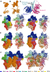
Figure S7.. Additional data together with Fig 7 showing the Noc3p, Noc3p dimer, ORC-DNA, ORC-Noc3p-DNA, and (ORC-Noc3p)2 dimer–DNA models.
(A, B, C, D, E, F) Modeling was performed manually (B, D, E, F) or by using HADDOCK2.2 (C), with the published Noc3p (A) and ORC (D) cryo-EM structures as inputs. Ribbon diagrams and space-filling models are provided. (D, E) Views from two angles are shown in (D, E). (A) Published Noc3p monomer structure. (B) Noc3p dimer model. (C) Noc3p dimer model built by using HADDOCK2.2. Our model indicates that Noc3p can form a dimer through the central helix. Noc3p is composed of a helix bundle in which a long and central helix is formed by residues 370–420 aa. The terminals, monomers, and structures are as indicated. (D) Published model of ORC single-hexamer with DNA. (E) Model of ORC single-hexamer with Noc3p and DNA. (F) Model of (ORC-Noc3p)2 dimer with DNA.
Figure 7.
Figure 7.. Models for (Noc3p-ORC)2 dimerization, pre-RC formation, and (Noc3p-ORC)2 de-dimerization.
Free (non–chromatin-bound) Noc3p-ORC binds to the origin-bound Noc3p-ORC at each replication origin by protein–protein interactions to form a (Noc3p-ORC)2 dimer at the M-to-G1 transition to enable loading of the MCM double-hexamer. Upon replication initiation, each (Noc3p-ORC)2 dimer de-dimerizes and separates into two Noc3p-ORCs, which bind and protect the two nascent replication origins at each replication bubble (See Discussion section for details).

References

    1. Amin A, Cheung MH, Liang C (2019) DNA replication-initiation proteins in eukaryotic cells. Biomed J Sci Tech Res 23: 17042–17049. 10.26717/BJSTR.2019.23.003830 - DOI
    1. Amin A, Wu R, Cheung MH, Scott JF, Wang Z, Zhou Z, Liu C, Zhu G, Wong CKC, Yu Z, et al. (2020) An essential and cell cycle-dependent ORC dimerization cycle regulates eukaryotic chromosomal DNA replication. Cell Rep 30: 3323–3338.e6. 10.1016/j.celrep.2020.02.046 - DOI - PubMed
    1. Bassler J, Kallas M, Hurt E (2006) The NUG1 GTPase reveals an N-terminal RNA-binding domain that is essential for association with 60 S pre-ribosomal particles. J Biol Chem 281: 24737–24744. 10.1074/jbc.m604261200 - DOI - PubMed
    1. Bell SP, Labib K (2016) Chromosome duplication in Saccharomyces cerevisiae. Genetics 203: 1027–1067. 10.1534/genetics.115.186452 - DOI - PMC - PubMed
    1. Ben-Aroya S, Coombes C, Kwok T, O’Donnell KA, Boeke JD, Hieter P (2008) Toward a comprehensive temperature-sensitive mutant repository of the essential genes of Saccharomyces cerevisiae. Mol Cell 30: 248–258. 10.1016/j.molcel.2008.02.021 - DOI - PMC - PubMed

Publication types

MeSH terms

Substances

LinkOut - more resources