Review
. 2023 Mar;33(2):e13147.
doi: 10.1111/bpa.13147. Epub 2023 Jan 4.

The neurovasculature as a target in temporal lobe epilepsy

Affiliations
Review

The neurovasculature as a target in temporal lobe epilepsy

Yvonne Reiss et al. Brain Pathol. 2023 Mar.

Abstract

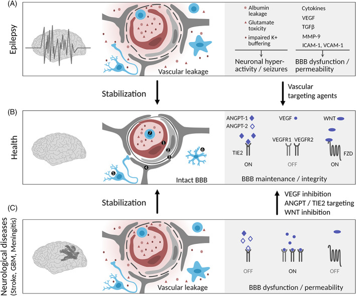
The blood-brain barrier (BBB) is a physiological barrier maintaining a specialized brain micromilieu that is necessary for proper neuronal function. Endothelial tight junctions and specific transcellular/efflux transport systems provide a protective barrier against toxins, pathogens, and immune cells. The barrier function is critically supported by other cell types of the neurovascular unit, including pericytes, astrocytes, microglia, and interneurons. The dysfunctionality of the BBB is a hallmark of neurological diseases, such as ischemia, brain tumors, neurodegenerative diseases, infections, and autoimmune neuroinflammatory disorders. Moreover, BBB dysfunction is critically involved in epilepsy, a brain disorder characterized by spontaneously occurring seizures because of abnormally synchronized neuronal activity. While resistance to antiseizure drugs that aim to reduce neuronal hyperexcitability remains a clinical challenge, drugs targeting the neurovasculature in epilepsy patients have not been explored. The use of novel imaging techniques permits early detection of BBB leakage in epilepsy; however, the detailed mechanistic understanding of causes and consequences of BBB compromise remains unknown. Here, we discuss the current knowledge of BBB involvement in temporal lobe epilepsy with the emphasis on the neurovasculature as a therapeutic target.

Keywords: VEGF/VEGFR; Wnt/β-catenin; angiopoietin/Tie2; blood-brain barrier; cerebral edema; epilepsy animal models; in vitro BBB models; neurovascular unit; temporal lobe epilepsy.


Conflict of interest statement

The authors declare no competing interest.

Figures

FIGURE 1
FIGURE 1
Vascular dysfunction at the neurovascular unit in epilepsy. Schematic drawing of the cellular and molecular composition of the NVU in health, epilepsy, and neurological diseases with BBB dysfunction (stroke, GBM, meningitis). (A) In epilepsy, BBB disruption leads to leakage of blood components (albumin, neurotransmitter (e.g., glutamate), K+ ions), leukocyte infiltration, aberrant transport and clearance of molecules, neuronal hyperactivity, and BBB damage. Microglia and astrocytes release cytokines and growth factors that further promote EC activation and BBB dysfunction. (B) An intact BBB protects the brain parenchyma from factors present in the systemic circulation and maintains a highly regulated brain micromilieu required for brain homeostasis and proper neuronal functioning. (C) BBB integrity can be re‐established upon vascular‐targeting therapies in neurological diseases associated with BBB dysfunction and cerebral edema. Endothelial cell (1), pericyte (2), basal lamina (3), astrocyte (4), neuron (5), microglia/activated microglia (6), leukocyte (7). Illustration: Visual Science Communication
FIGURE 2
FIGURE 2
Schematic of the physiological underpinnings of postictal imaging findings and presentation of exemplary cases. Patient I: 52 years old, male, no lesion in MRI, focal seizure with impaired awareness and motor onset, ictal EEG onset in left fronto‐temporal electrodes. Postictal imaging finding: Left frontal cortical T2 hyperintensity, hypointense thickened cortex in T1, hyperintense diffusion signal. Patient II: 34 years old, male, resected left‐temporal cavernoma, generalized tonic–clonic seizure, ictal EEG onset in left centro‐temporal electrodes. Postictal imaging finding: Postictally reduced qT1 as compared to the interictal volume indicative of Gd+‐accumulation. Patient III: 27 years old, female, no lesion in MRI, focal seizure with impaired awareness and motor onset, no EEG correlate. Postictal imaging finding: Postictal hyperperfusion in right precuneus as comparted to interictal SPECT volume. DWI, diffusion weighted image; Gd+, gadolinium; ISAS: ictal‐interictal SPECT analyzed by SPM; qT1, quantitative T1; T2w, T2 weighted image; T1w, T1 weighted image; SPECT, single photon emission computed tomography; ΔT1, interictal qT1—postictal qT1
FIGURE 3
FIGURE 3
In vitro BBB co‐culture model of hippocampal slices with brain endothelial cells. (A) Brain endothelial cells (EC) isolated from the temporal cortex surgically resected from TLE patients by selective amygdalahippocampectomy (top panel; adapted from [139]) exhibit the classic spindle shape of brain ECs (middle panel) and express the BBB markers VE‐cadherin (white) and claudin‐5 (red). Scale bars 10 mm. (B) The isolated brain ECs cultured on transwell inserts represent a BBB in vitro monoculture model. The co‐culture of the cortical ECs on the transwell insert membrane (apical) with the hippocampal tissue slice obtained from TLE patient surgery in the bottom chamber (basal) represent the non‐contact co‐culture model for the BBB in vitro. The co‐culture of the hippocampal slice on top of the transwell insert membrane (basal) with the cortical ECs on the bottom of the insert membrane (apical) represents a contact co‐culture BBB in vitro epilepsy model. Impedance measurements performed on these transwell inserts for the in vitro BBB model in a cellZscope (nanoAnalytics) device provide resistance and capacitance values reflecting BBB function. Electrophysiological recordings from the hippocampal slice either from the insert or from the bottom chamber provide measurements of seizure‐like events from the epileptic tissue. (C) Representative transendothelial electrical resistance (TEER) and capacitance (Ccl) values from a monolayer of cortical brain ECs (green line) isolated from a TLE patient show high resistance and low capacitance values indicating a functional monoculture BBB model in vitro (as in A) compared to the cell‐free insert (black line).

References

    1. Thijs RD, Surges R, O'Brien TJ, Sander JW. Epilepsy in adults. Lancet. 2019;393(10172):689–701. - PubMed
    1. Rosenow F, Lüders H. Presurgical evaluation of epilepsy. Brain. 2001;124(9):1683–700. - PubMed
    1. Willems LM, Hamer HM, Knake S, Rosenow F, Reese JP, Strzelczyk A. General trends in prices and prescription patterns of anticonvulsants in Germany between 2000 and 2017: analysis of national and cohort‐based data. Appl Health Econ Health Policy. 2019;17(5):707–22. - PubMed
    1. Tang F, Hartz AMS, Bauer B. Drug‐resistant epilepsy: multiple hypotheses, few answers. Front Neurol. 2017;8:301. - PMC - PubMed
    1. Strzelczyk A, Griebel C, Lux W, Rosenow F, Reese JP. The burden of severely drug‐refractory epilepsy: a comparative longitudinal evaluation of mortality, morbidity, resource use, and cost using German health insurance data. Front Neurol. 2017;8:712. - PMC - PubMed

Publication types