. 2023 Jan;613(7943):383-390.
doi: 10.1038/s41586-022-05565-5. Epub 2023 Jan 4.

Structures and mechanisms of tRNA methylation by METTL1-WDR4

Affiliations

Structures and mechanisms of tRNA methylation by METTL1-WDR4

Victor M Ruiz-Arroyo et al. Nature. 2023 Jan.

Abstract

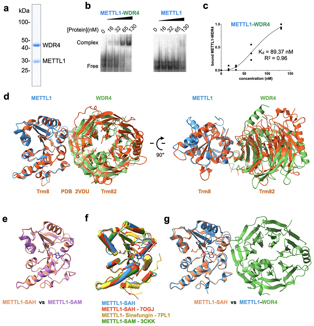
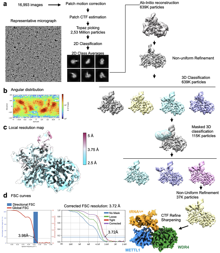
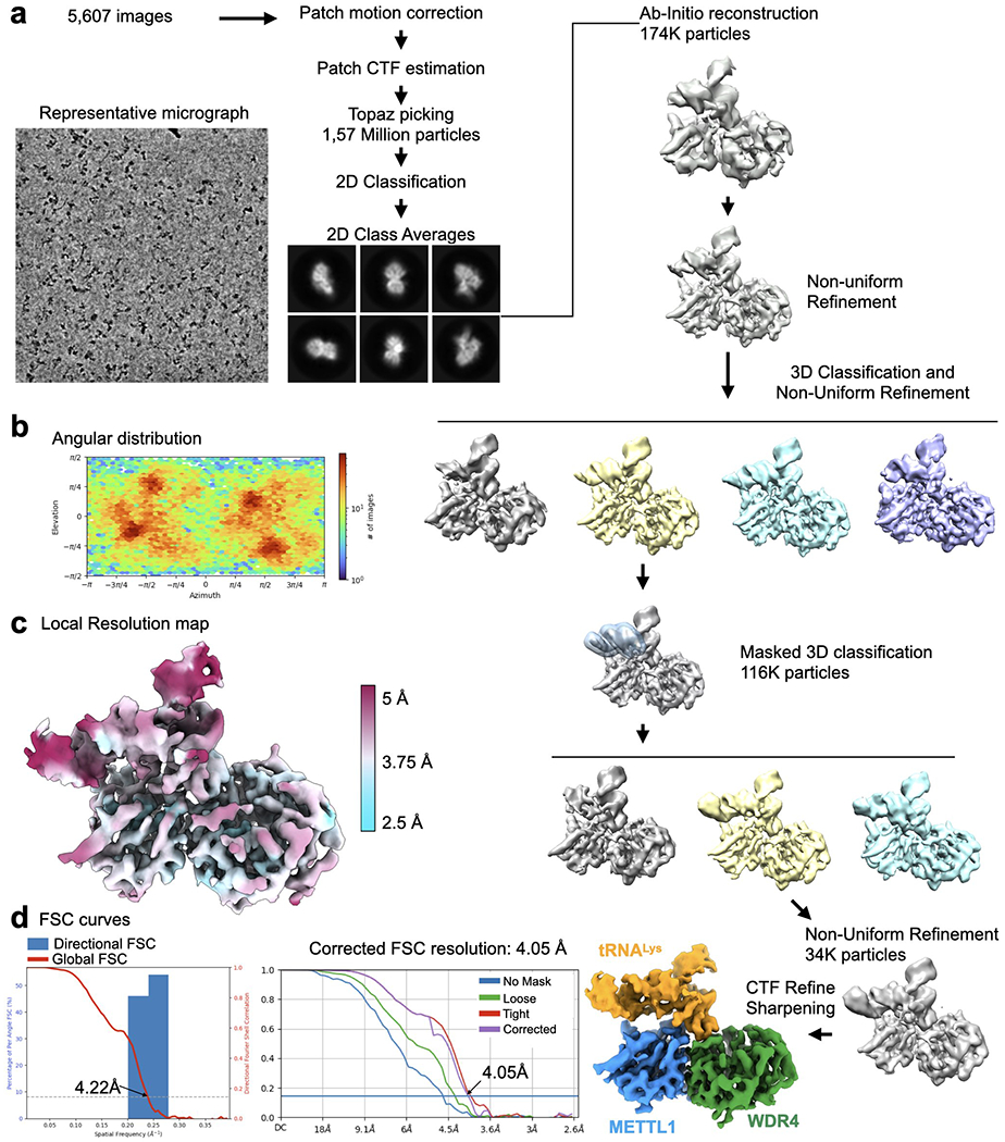
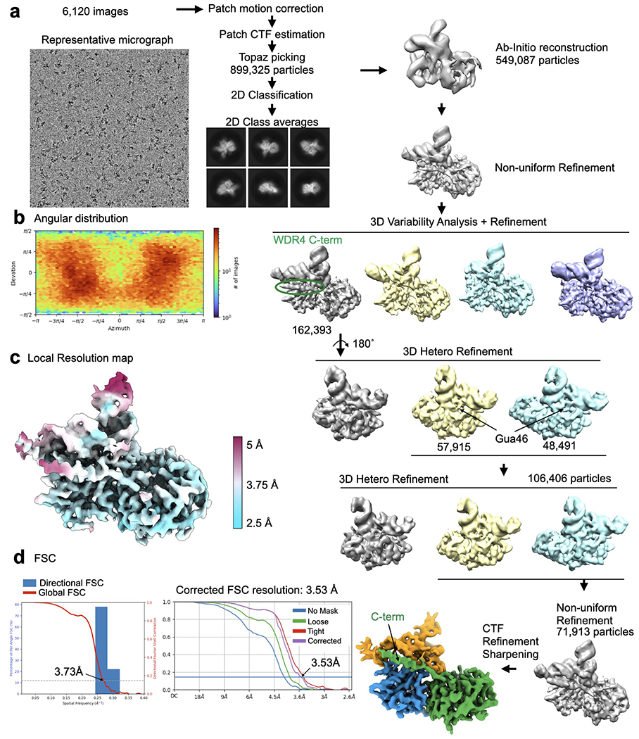
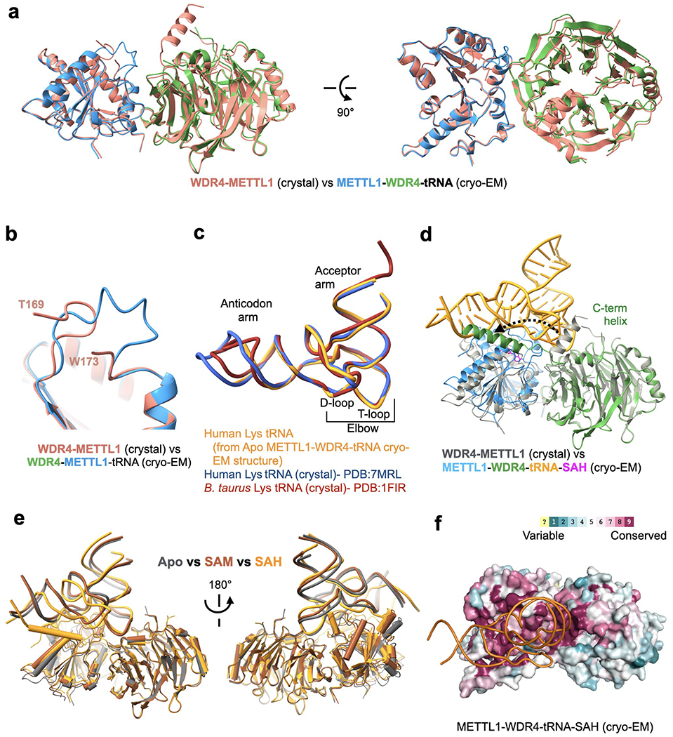
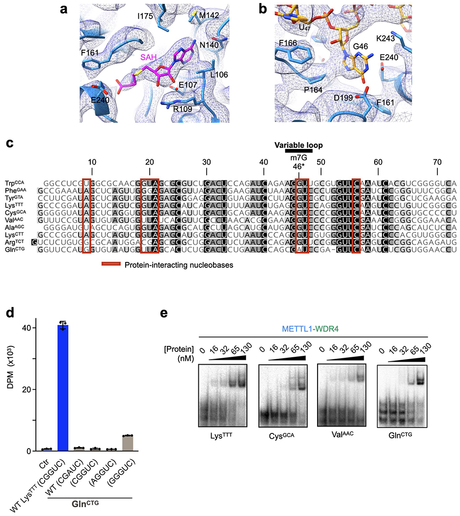
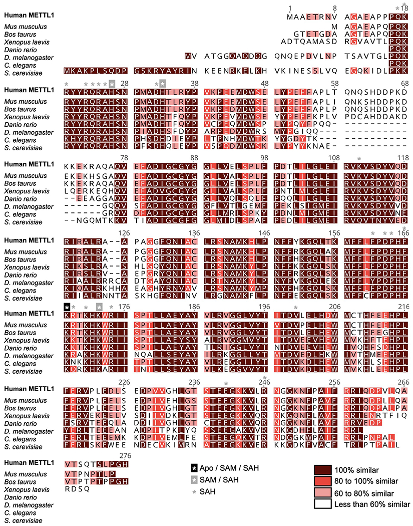
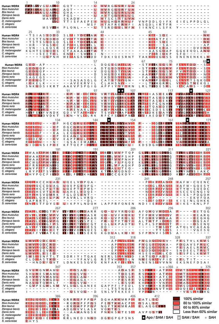
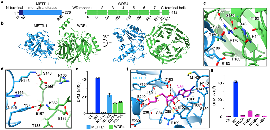
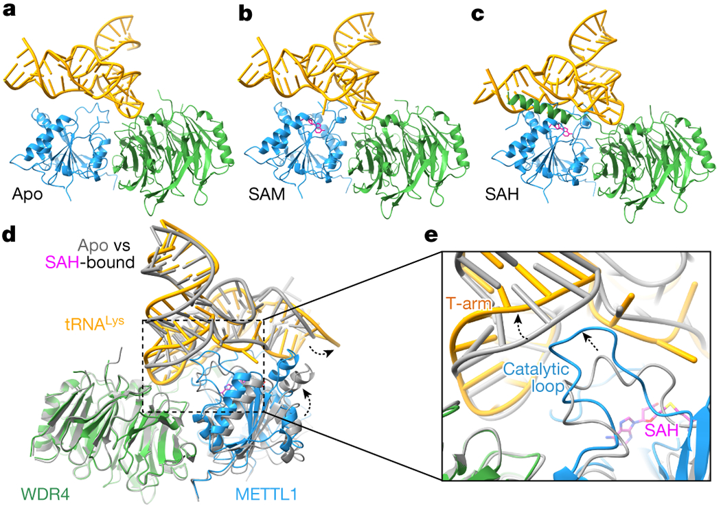
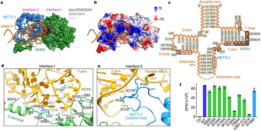
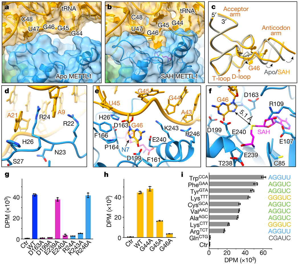
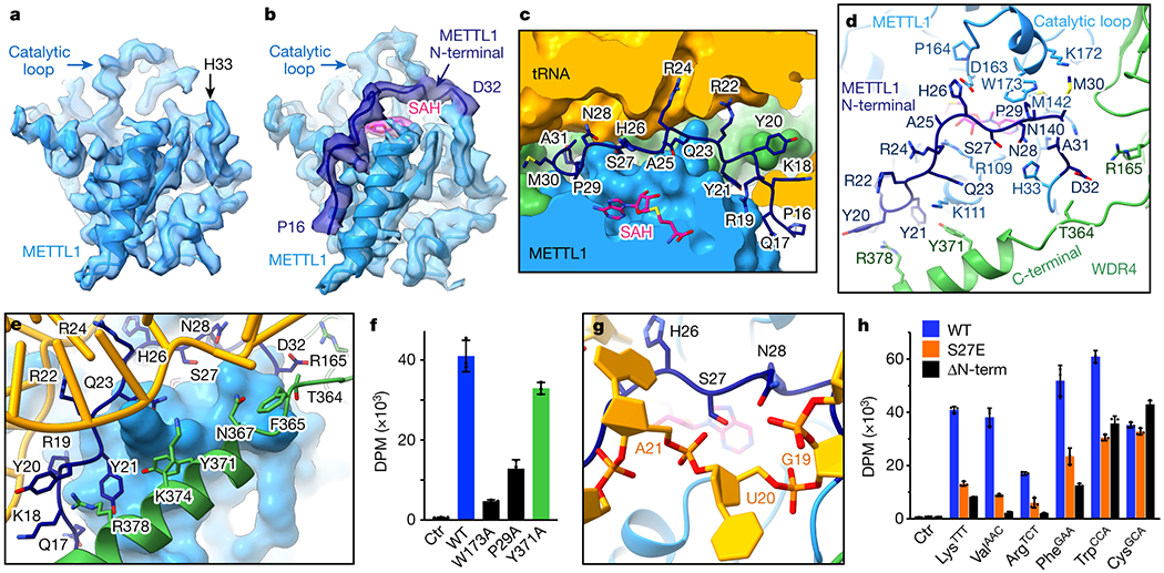
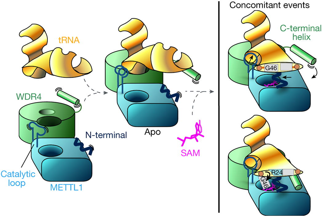
Specific, regulated modification of RNAs is important for proper gene expression1,2. tRNAs are rich with various chemical modifications that affect their stability and function3,4. 7-Methylguanosine (m7G) at tRNA position 46 is a conserved modification that modulates steady-state tRNA levels to affect cell growth5,6. The METTL1-WDR4 complex generates m7G46 in humans, and dysregulation of METTL1-WDR4 has been linked to brain malformation and multiple cancers7-22. Here we show how METTL1 and WDR4 cooperate to recognize RNA substrates and catalyse methylation. A crystal structure of METTL1-WDR4 and cryo-electron microscopy structures of METTL1-WDR4-tRNA show that the composite protein surface recognizes the tRNA elbow through shape complementarity. The cryo-electron microscopy structures of METTL1-WDR4-tRNA with S-adenosylmethionine or S-adenosylhomocysteine along with METTL1 crystal structures provide additional insights into the catalytic mechanism by revealing the active site in multiple states. The METTL1 N terminus couples cofactor binding with conformational changes in the tRNA, the catalytic loop and the WDR4 C terminus, acting as the switch to activate m7G methylation. Thus, our structural models explain how post-translational modifications of the METTL1 N terminus can regulate methylation. Together, our work elucidates the core and regulatory mechanisms underlying m7G modification by METTL1, providing the framework to understand its contribution to biology and disease.


Conflict of interest statement

Competing Interests

The authors declare no competing interests.

Figures

Extended Data Fig. 1:
Extended Data Fig. 1:. METTL1-WDR4 protein purification and tRNA complex reconstitution.
a, SDS-PAGE of purified full-length wild-type METTL1-WDR4 complex. A representative gel among 3 replicates is shown. b, EMSA to measure the affinity for tRNALys. For each gel, protein concentrations are 0, 16, 32, 65, and 130 nM, left to right. Representative gel among 3 replicates is shown. c, Quantification of EMSA for METTL1-WDR4 from 3 replicate experiments. d, Superimposition of yeast Trm8-Trm82 (PDB 2VDU, orange) onto the crystal structure of human METTL1-WDR4. The complex structures were superimposed by aligning METTL1 with Trm8. e-g, Superimposition of METTL1 structures as indicated. The structures missing PDB codes are from this study.
Extended Data Fig. 2:
Extended Data Fig. 2:. Cryo-EM data processing for the METTL1-WDR4-tRNALys structure
a, Cryo-EM data processing workflow. Left, a representative micrograph of the METTL1-WDR4-tRNA complex particles. A total of 16,993 images were used for picking and 2D classification. 2D class averages that showed high-resolution features were used for further 3D analysis using Cryosparc. 3D classification identified different populations that showed partial or no density around the anticodon arm. 3D classification applying the indicated mask revealed a homogeneous population of particles showing contiguous density in the tRNA region. b, Angular distribution plot. c, Local resolution map shown with colors on the sharpened map. d, Directional FSC plot and FSC curves showing the resolution at 0.143 cutoff.
Extended Data Fig. 3:
Extended Data Fig. 3:. Cryo-EM data processing for the METTL1-WDR4-tRNALys-SAM structure.
a, Cryo-EM data processing workflow. Left, a representative micrograph of the METTL1-WDR4-tRNA-SAM complex particles. A total of 5,607 images were used for picking and 2D classification. 2D class averages that showed high-resolution features were used for further 3D analysis using Cryosparc. 3D classification identified different populations that showed partial density around the anticodon arm. 3D classification applying the indicated mask revealed a homogeneous population of particles showing contiguous density in the tRNA region. b, Angular distribution plot. c, Local resolution map shown with colors on the sharpened map. d, Directional FSC plot and FSC curves showing the resolution at 0.143 cutoff.
Extended Data Fig. 4:
Extended Data Fig. 4:. Cryo-EM data processing for the METTL1-WDR4-tRNALys-SAH structure.
a, Cryo-EM data processing workflow. Left, a representative micrograph of the METTL1-WDR4-tRNA-SAH complex particles. A total of 6,120 images were used for picking and 2D classification. 2D class averages that showed high-resolution features were used for further 3D analysis using Cryosparc. 3D variability analysis identified a population of particles that showed consistent density for the WDR4 C-terminal helix and several rounds of 3D Heterogeneous Refinement identified particles showing prominent density for G46. b, Angular distribution plot. c, Local resolution map shown with colors on the sharpened map. d, Directional FSC plot and FSC curves showing the resolution at 0.143 cutoff.
Extended Data Fig. 5:
Extended Data Fig. 5:. Conformational changes in structures containing the METTL1-WDR4-tRNA complex.
a, Superimposition of the METTL1-WDR4 crystal structure on the METTL1-WDR4-tRNA cryo-EM structure, aligned by METTL1. b, Flexible loop of METTL1 (161-175, catalytic loop) becomes more ordered with RNA. c, Superimposition of different tRNALys structures. All three have the same sequence except the tips of the anticodon arm and the acceptor arm. The B. Taurus structure (PDB:1FIR) was with a fully modified tRNA and the other two structures were obtained for unmodified RNA after in vitro transcription. d, Superimposition of the METTL1-WDR4 crystal structure (gray) onto the SAH-bound quaternary complex cryo-EM structure (multiple colors), aligned by WDR4. Movement of the WDR4 C-terminal helix upon binding RNA is shown with a dashed arrow. e, Superimposition of all three cryo-EM structures (colored by state) presented in this study, aligned by WDR4. f. Surface representation of the SAH-bound cryo-EM structure colored by evolutionary sequence conservation (Consurf server). The orientation is identical to Fig. 3a and 3b.
Extended Data Fig. 6:
Extended Data Fig. 6:. Structural and sequence organization of tRNA.
a-b, Sharpened cryo-EM map (mesh) near the SAH-binding site (a) and the G46 binding pocket (b). c, Sequence alignment of the human tRNAs used in this study shaded by conservation. Red boxes indicate nucleobases within 4 Å of protein. Sequences were aligned using Clustal Omega and visualized by Geneious Prime. d, In vitro methylation activity of full-length METTL1-WDR4 for the indicated tRNAs with the specified variable loop sequences, shown as mean ± SD from 3 replicates. e, EMSA using METTL1-WDR4 with different tRNAs shows no dramatic differences in affinities. Representative images from 3 replicate experiments are shown. For each gel, protein concentrations are 0, 16, 32, 65, and 130 nM, left to right.
Extended Data Fig. 7:
Extended Data Fig. 7:. Sequence alignment of METTL1 protein homologs.
Sequences used are from human (Q9UBP6), Mus musculus (Q9Z120), Bos taurus (Q2YDF1), Xenopus laevis (Q6NU94), Danio rerio (Q5XJ57), Drosophila melanogaster (O77263), Caenorhabditis elegans (Q23126) and Saccharomyces cerevisiae (Q12009) METTL1. Sequences were aligned using Clustal Omega and visualized by Geneious Prime. Residues within 4 Å of RNA in different states are indicated with asterisks.
Extended Data Fig. 8:
Extended Data Fig. 8:. Sequence alignment of WDR4 protein homologs.
Sequences used are from human (P57081), Mus musculus (Q9EP82), Bos taurus (A7E3S5), Xenopus laevis (Q7ZY78), Danio rerio (A4IGH4), Drosophila melanogaster (Q9W415), Caenorhabditis elegans (Q23232) and Saccharomyces cerevisiae (A6ZYC3) WDR4. Sequences were aligned using Clustal Omega and visualized by Geneious Prime. Residues within 4 Å of RNA in different states are indicated with asterisks.
Fig. 1:
Fig. 1:. Architecture of the human METTL1-WDR4 complex.
a, Domain organization of human METTL1 and WDR4. b, Crystal structure of the METTL1-WDR4 complex. c-d, Close-up views of the METTL1-WDR4 interface. R170 interactions with nearby backbone carbonyls build a scaffold to enable nearby hydrophobic intermolecular interactions. Side chains within 4 Å of the other protein or R170 of WDR4 are shown. Hydrogen bonds are indicated with dashed lines. e, In vitro methylation activity of full-length METTL1-WDR4 with indicated point mutations, shown as mean ± SD from 3 replicates. f, Close-up view of the SAH-binding pocket in the crystal structure of METTL1. Side chains are shown as sticks for the residues within 4 Å of SAH and waters as red spheres. Dashed lines indicate hydrogen bonds. g, In vitro methylation activity of the full-length METTL1-WDR4 complex with indicated point mutations in METTL1, shown as mean ± SD from 3 replicates.
Fig. 2:
Fig. 2:. Architecture of the METTL1-WDR4-tRNA complex in three states.
a-c, Models of the cryo-EM structures in the indicated cofactor binding states, in cartoon representation. METTL1, WDR4, tRNA and SAM/SAH are colored blue, green, gold, and magenta, respectively. d, Superimposition of the apo and SAH-bound cryo-EM structures of METTL1-WDR4-tRNA, aligning by WDR4. METTL1 moves closer to tRNA in the presence of SAH (black dashed arrow). e, Zoomed-in view of (d) showing the tRNA and the catalytic loop protruding toward the tRNA (black dashed arrow).
Fig. 3:
Fig. 3:. Structural recognition of tRNA shape by METTL1-WDR4.
a, Surface representation of the METTL1-WDR4-tRNA-SAH structure, with only tRNA in ribbon representation. Residues within 4 Å of RNA are colored by conservation of the protein-RNA contact in the three states: black (Apo/SAM/SAH), grey (SAM/SAH), or white (SAH only). Two interface areas with conserved contacts in all three states are marked with dotted rectangles in magenta. b, Vacuum electrostatics surface representation of the protein complex in the same orientation as (a). c, Secondary structure diagram of tRNALys. Nucleotides within 4 Å of protein are indicated with rounded squares and colored using the same color key as in (a). d-e, Close-up views of METTL1-WDR4-tRNA-SAH where the residues within 4 Å of RNA are shown with side chain sticks for the interfaces marked in (a). f, In vitro methylation activity of METTL1-WDR4 with point mutations in WDR4 (green) or METTL1 (blue), shown as mean ± SD from 3 replicates.
Fig. 4:
Fig. 4:. Detailed view of the active site.
a-b, View of the tRNA variable near METTL1. RNA (yellow) and METTL1 (blue) are shown with transparent surface representation and cartoon, and SAH is shown in magenta. The variable loop is indicated with nucleotide numbers. The gap in apo state is filled as G46 is released from the helical stack and buried in the catalytic pocket after binding SAH. The orientation is the same as in (a). c. Twisting near the variable loop exposes G46 and rotates the anticodon arm relative to the rest of the tRNA. d, R24 of METTL1 occupies the space left behind by G46 to stack between A21 and A9. e, View of G46 base surrounded by a triad of acidic residues next to SAH. N7 position is indicated with an arrow, and side chains are shown only for the residues within 4 Å of RNA. f, Distance between the sulfur in SAH and N7 in G46 is indicated with an arrow. g-i, In vitro methylation activity of METTL1-WDR4 for tRNALys with point mutations in METTL1 (g) or tRNALys (h), or with wild-type proteins and different human tRNAs (i). All bar graphs are shown as mean ± SD from 3 replicates.
Fig. 5:
Fig. 5:. METTL1 N-term coordinates SAH binding with RNA and protein conformational changes.
a-b, Sharpened cryo-EM maps of METTL1-WDR4-tRNA ± SAH structures reveal that the METTL1 N-terminal peptide (dark blue) becomes ordered upon binding SAH. c, METTL1 N-term and SAH shown in stick representation with surrounding proteins and RNA shown as surfaces. N-term seals the gap in the protein-RNA interface. d, METTL1 N-term supports conformational changes in the catalytic loop and the WDR4 C-terminal helix. Side chains within 4 Å of the N-term are shown in stick representation. e, METTL1 N-term and WDR4 C-terminal helix support each other to bind RNA together. Side chains within 4 Å of RNA or N-term are shown with sticks. f, In vitro methylation activity of full-length METTL1-WDR4 constructs with point mutations in WDR4 (green) or METTL1 (dark blue), shown as mean ± SD from 3 replicates. g, Close-up view of METTL1 S27 lying on top of SAH and under the RNA backbone. h, In vitro methylation activity of full-length METTL1-WDR4, using WT, S27E, or N-terminally truncated (Δ1-19) METTL1. Data are shown as mean ± SD from 3 replicates.
Fig. 6:
Fig. 6:. Mechanistic model for tRNA m7G46 methylation by METTL1-WDR4.
The stable heterodimeric METTL1-WDR4 protein complex provides a docking site for the tRNA Elbow. The protein-RNA docking can occur without cofactor, and METTL1 can bind SAM without tRNA. In the apo state without SAM or SAH, the catalytic loop becomes more ordered with bound RNA. WDR4 C-terminal helix moves close to RNA but remains flexible and METTL1 N-term is also disordered. When both tRNA and SAM are bound, METTL1 shifts even closer to the tRNA—the catalytic loop protrudes toward the tRNA, METTL1 N-term becomes ordered sandwiched between RNA and SAM, and WDR4 C-term attaches to the METTL1 N-term to stabilize the bound RNA together. The same N-term also supports the twisting of the tRNA to release G46 by replacing the stacking interactions with R24.

References

    1. Frye M, Harada BT, Behm M & He C RNA modifications modulate gene expression during development. Science 361, 1346–1349 (2018). - PMC - PubMed
    1. Wiener D & Schwartz S The epitranscriptome beyond m(6)A. Nat Rev Genet 22, 119–131 (2021). - PubMed
    1. Pan T Modifications and functional genomics of human transfer RNA. Cell Res 28, 395–404 (2018). - PMC - PubMed
    1. Suzuki T The expanding world of tRNA modifications and their disease relevance. Nat Rev Mol Cell Biol 22, 375–392 (2021). - PubMed
    1. Alexandrov A, Martzen MR & Phizicky EM Two proteins that form a complex are required for 7-methylguanosine modification of yeast tRNA. RNA 8, 1253–1266 (2002). - PMC - PubMed

References for Methods

    1. Studier FW Protein production by auto-induction in high density shaking cultures. Protein Expr Purif 41, 207–234 (2005). - PubMed
    1. Minor W, Cymborowski M, Otwinowski Z & Chruszcz M HKL-3000: the integration of data reduction and structure solution – from diffraction images to an initial model in minutes. Acta Crystallographica Section D: Biological Crystallography 62, 859–866 (2006). - PubMed
    1. McCoy AJ et al. Phaser crystallographic software. J Appl Crystallogr 40, 658–674 (2007). - PMC - PubMed
    1. Emsley P & Cowtan K Coot: model-building tools for molecular graphics. Acta Crystallogr D Biol Crystallogr 60, 2126–2132 (2004). - PubMed
    1. Adams PD et al. PHENIX: a comprehensive Python-based system for macromolecular structure solution. Acta Crystallogr D Biol Crystallogr 66, 213–221 (2010). - PMC - PubMed

Publication types

MeSH terms

LinkOut - more resources