. 2023 Jan;613(7943):391-397.
doi: 10.1038/s41586-022-05566-4. Epub 2023 Jan 4.

Structural basis of regulated m7G tRNA modification by METTL1-WDR4

Affiliations

Structural basis of regulated m7G tRNA modification by METTL1-WDR4

Jiazhi Li et al. Nature. 2023 Jan.

Abstract

Chemical modifications of RNA have key roles in many biological processes1-3. N7-methylguanosine (m7G) is required for integrity and stability of a large subset of tRNAs4-7. The methyltransferase 1-WD repeat-containing protein 4 (METTL1-WDR4) complex is the methyltransferase that modifies G46 in the variable loop of certain tRNAs, and its dysregulation drives tumorigenesis in numerous cancer types8-14. Mutations in WDR4 cause human developmental phenotypes including microcephaly15-17. How METTL1-WDR4 modifies tRNA substrates and is regulated remains elusive18. Here we show, through structural, biochemical and cellular studies of human METTL1-WDR4, that WDR4 serves as a scaffold for METTL1 and the tRNA T-arm. Upon tRNA binding, the αC region of METTL1 transforms into a helix, which together with the α6 helix secures both ends of the tRNA variable loop. Unexpectedly, we find that the predicted disordered N-terminal region of METTL1 is part of the catalytic pocket and essential for methyltransferase activity. Furthermore, we reveal that S27 phosphorylation in the METTL1 N-terminal region inhibits methyltransferase activity by locally disrupting the catalytic centre. Our results provide a molecular understanding of tRNA substrate recognition and phosphorylation-mediated regulation of METTL1-WDR4, and reveal the presumed disordered N-terminal region of METTL1 as a nexus of methyltransferase activity.


Conflict of interest statement

Competing interests

R.I.G. is a co-founder and scientific advisory board member of 28/7 Therapeutics and Theonys. E.S.F. is a founder, science advisory board (SAB) member, and equity holder in Civetta Therapeutics, Lighthorse Therapeutics, Neomorph Inc (board of directors), and Proximity Therapeutics. SAB member and equity holder in Avilar Therapeutics and Photys Therapeutics. E.S.F. is a consultant to Novartis, Sanofi, EcoR1 capital, Avilar, and Deerfield. The Fischer lab receives or has received research funding from Astellas, Novartis, Voronoi, Interline, Ajax, and Deerfield. The rest of the authors declare no competing interests.

Figures

Extended Data Fig. 1 |
Extended Data Fig. 1 |. Sample preparation and quality check.
(a-b) tRNA candidates for ternary complex reconstitution. Schematic representations of yeast tRNAPhe and human tRNAVal. Yeast tRNAPhe is a matured tRNAPhe purified from yeast (Sigma) (a). Human tRNAVal is an annealed single strand RNA oligos synthesized based on human tRNAVal-TAC sequence (Horizon) (b). (c) Chromatography traces and SDS-PAGE analysis of purified METTL1-WDR4 complex (absorption at 280nm and 254nm). For gel source data, see Supplementary Fig. 3. (d) Gel filtration profiles of free tRNAPhe (blue), METTL1-WDR4 binary complex (green), METTL1-WDR4-tRNAPhe ternary complex (purple) and METTL1-WDR4-tRNAVal ternary complex (yellow) (absorption at 280nm are shown). (e) Identification of the reconstituted METTL1-WDR4-tRNAPhe ternary complex. Sample in the main peak of the reconstitution chromatography trace is analyzed by native PAGE. tRNA and protein are virtualized by EB and G250 staining separately. For gel source data, see Supplementary Fig. 4.
Extended Data Fig. 2 |
Extended Data Fig. 2 |. Cryo-EM workflows and the quality of reconstructed cryo-EM maps.
(a-b) Workflow of 3D reconstruction of METTL1-WDR4-tRNAPhe dataset (a) and the METTL1-WDR4-tRNAVal dataset (b). (c-d) Fourier shell correlation (FSC) curves (upper panel) and orientation distributions (lower panel) of 3D reconstructed METTL1-WDR4-tRNAPheand METTL1-WDR4-tRNAVal cryo-EM maps. See also Extended Data Table 1. (e) Histogram of directional FSC curves of METTL1-WDR4-tRNAPhe dataset (upper panel) and the METTL1-WDR4-tRNAVal dataset (lower panel). (f) Local resolutions of METTL1-WDR4-tRNAPhe (upper panel) and METTL1-WDR4-tRNAVal (lower panel) cryo-EM maps. (g) Representative segments of sharpened cryo-EM map fitted with the model. (h) State A and State B models of METTL1-WDR4-tRNAPhe fit in sharpened cryo-EM maps. The variable loop of tRNA fitted with sharpened (top) and unsharpened (bottom) maps from 3D variability analysis are shown in parallel with the whole model.
Extended Data Fig. 3 |
Extended Data Fig. 3 |. Similar binding mode of METTL1-WDR4 to tRNAPhe and tRNAVal.
(a) Cryo-EM density maps of METTL1-WDR4-tRNAVal complex and the corresponding atomic model. The unsharpened (left), sharpened (middle) and DeepEMhancer processed (right) density maps are shown. (b) Overall structural superposition of METTL1-WDR4-tRNAVal (slate) and METTL1-WDR4-tRNAPhe (white). (c) Electrostatic potential of METTL1 and WDR4 in ternary complex (tRNAPhe). Red, negative; blue, positive. Figure was generated using PyMOL. tRNA domains are colored according to Extended Data Fig. 1a. (d) Tilted loading of tRNAPhe onto the METTL1-WDR4 complex. Overall structure of METTL1-WDR4-tRNAPhe complex from the WDR4 side. The angle between the short axis of METTL1-WDR4 and the aminoacyl-branch axis of tRNA is about 130 degrees. The angle is measured utilizing residue 213 (WDR4), residue 163 (WDR4) and base 73 of tRNA using PyMOL.
Extended Data Fig. 4 |
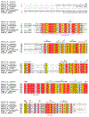
Extended Data Fig. 4 |. Sequence alignment of METTL1 proteins.
The human METTL1 protein sequence was aligned with its respective homologs. The secondary structure diagram for human METTL1 is shown on the top. Conserved residues are shaded in yellow, whereas essentially invariant residues are shown in red. The conserved N-terminal region, aC and a6 helix are underlined. K143 (key residue that interacts with WDR4) is highlighted with a blue star on the bottom. The alignment is performed with the Clustal Omega multiple sequence alignment program (EMBL-EBI) and visualized by ESPript 3.0 server.
Extended Data Fig. 5.
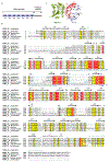
Extended Data Fig. 5.. Domain organization and sequence alignment of WDR4 proteins.
(a) Schematic representation of full-length WDR4 domains based on sequence, secondary structure prediction and experimental structures. WD, WD family repeat domain and are numbered with B1-B7. (b) Sequence alignment of WDR4 proteins. The secondary structure diagram (black, based on experimental structure; pale green, based on AlphaFold prediction) for human WDR4 is shown on the top. Conserved residues are shaded in yellow, whereas essentially invariant residues are shown in red. B3 and B4 are underlined in green and blue, respectively. Key residues are highlighted with stars on the bottom. Orange stars, residues involved in tRNA (T-arm) binding; magenta stars, patient related mutagenesis sites; blue stars, METTL1 interaction sites. The alignment is performed with the Clustal Omega multiple sequence alignment program (EMBL-EBI) and visualized by ESPript 3.0 server. (c) Structure of METTL1-WDR4-tRNAPhe ternary complex with top view (only METTL1 and WDR4 are shown). The conserved region of B2-B5 (WDR4) is highlighted in red.
Extended Data Fig. 6 |
Extended Data Fig. 6 |. The conformational change of METTL1-WDR4 upon tRNA binding.
(a)Structure comparison between METTL1-WDR4-tRNAPhe and METTL1-WDR4. The structures are superposed on WDR4 protein. The tRNA in the ternary complex is not shown for a better view. METTL1-WDR4-tRNAPhe, pink; METTL1-WDR4, Cyan. The α1, α2, α5 and α6 helices significantly shift toward WDR4 and tRNA side. Structural changes of the residues 164-173 fragment of METTL1 are highlighted with a dash line box. (b) Superposition of tRNA-free (cyan) and tRNA bound (pink) states of METTL1. The structures are superposed on METTL1 protein and only only METTL1 are shown. The loop (residues 164-173) connecting α1 and the core fold of METTL1 forms the αC helix upon tRNA binding. (c) Binary complex model from METTL1-WDR4-tRNAPhe cryo-EM dataset fit in sharpened (top) and unsharpened (bottom) cryo-EM maps. (d) Structure comparison between EM_Binary (teal) and crystal METTL1-WDR4 (gray). The METTL1 protein is superimposed. (e) Local resolution of binary complex map. The αC loop of METTL1 region is highlighted with dashed line circle. (f)The αC loop of METTL1 (EM_Binary) fit in sharpened (top) and unsharpened (bottom) cryo-EM maps.
Extended Data Fig. 7 |
Extended Data Fig. 7 |. Essential residues of METTL1-WDR4 for MTase activity and tRNA recognition.
(a) Magnified view of METTL1-WDR4 interface in the crystal binary complex structure. The interactions of key residues are shown in dashed lines. K143 (METTL1) forms a salt bridge with D166 (WDR4); hydrogen bonds are formed between Y37 (METTL1) and E167 (WDR4), N147 (METTL1), and K168 (WDR4), K40 (METTL1) and mainchain of L185 (WDR4). (b) Relative methyltransferase activity of METTL1-WDR4 complexes expressed with indicated mutations. WT, wild type; Mut, catalytic dead double mutant (L160A/D163A). Two technical replicates were performed. (c-d) In vivo rescue experiment with METTL1 or WDR4 carrying indicated mutations in WDR4 (KO) cell lines (c) or METTL1 (KO) cell lines (d). n=2, biologically independent samples. Expression of WT and variants METTL1 or WDR4 is checked by western blot (lower panel). For gel source data, see Supplementary Fig. 5–6. (e) Structure comparison between METTL1-WDR4 crystal structure (PDB 7U20, METTL1, splitpea; WDR4, light blue) and Trm8-Trm82 (PDB 2VDU, orange). The WDR4 protein is superimposed. (f) Distance measurement between METTL1 E183 (OE2) and WDR4 R170 (NH1) in the crystal binary complex structure. 2Fo-Fc map is shown (1.2 σ). Figures were generated in Coot. (g, h) Western blot detection of overexpressed METTL1 or WDR4 in rescue experiments relative to Fig 2e, j. n=2, biologically independent samples. For gel source data, see Supplementary Fig. 7.
Extended Data Fig. 8 |
Extended Data Fig. 8 |. The METTL1 N-terminus plays important roles in catalytic regulation.
(a) Superimposed METTL1 N-terminus of available structures. The first visible residues of human METTL1 are labeled. (b) The IDDT value of the predicted METTL1-WDR4 structure. (c) The interactions between the N-terminal and α2 helix in AlphaFold prediction. (d) AlphaFold predicted METTL1 is superposed onto METTL1-WDR4-tRNAPhe. The predicted residues 16-21 insert into the space between METTL1 and tRNA. (e) Residue-specific secondary structure propensities derived from 1HN, 15N, 13C’, 13Cα and 13Cβ chemical shifts assignments. α-helix (red), coil/unstructured (grey), β-strand (green). (f) Competitive TR-FRET binding assay of labeled full-length METTL1-WDR4 with unlabeled proteins. The determined IC50 is listed. Two technical replicates were performed. (g) Western blot detection of overexpressed METTL1 or WDR4 relative to Fig. 3f. n=2, biologically independent samples. For gel source data, see Supplementary Fig. 7. (h) In vivo rescue experiment with indicated mutations in METTL1 (KO) cell lines (left). Expression of protein is checked by western blot (right). For gel source data, see Supplementary Fig. 8. (i) Key components of the G46 binding cavity fit in sharpened METTL1-WDR4-tRNAPhe cryo-EM map (mesh). Key residues and cofactor SAH are shown in stick. (j) Docking model of G46 flipping into the catalytic pocket with SAM bound. The transferred methyl group is indicated by arrow. Relevant elements are adjusted manually to make 180° angle and 2 Å between the guanine-N7 and S-CH3. The potential interactions between METTL1 and the base of G46 are highlighted with dashed lines. (k) Schematic diagram of the docking model depicting potential interactions (dashed lines) between G46 and SAM in the METTL1 active site prior to methyl transfer. (l-m) Relative methyltransferase activity of METTL1-WDR4 with buffer pH ranging from 5.6 to 8.33 (l) and indicated mutation (m). Two technical replicates were performed.
Fig. 1 |
Fig. 1 |. METTL1-WDR4 provides a platform for specific tRNA loading
(a-b) Cryo-EM density map of METTL1-WDR4-tRNAPhe complex and corresponding atomic model. The sharpened map is shown. METTL1, splitpea; WDR4, light blue; tRNAPhe, yellow; tRNA domains are colored according to Extended Data Fig. 1a. The G46 is highlighted in red. (c) Sail boat model of METTL1-WDR4-tRNAPhe ternary complex in side view (left) and front view (right). (d) X-ray crystal structure of full-length METTL1-WDR4 binary complex. METTL1 (splitpea) and WDR4 (light blue) are shown in cartoon representation. Secondary structure elements are numbered. (e) Structural comparison between METTL1-WDR4-tRNAPhe complex (pink) and METTL1-WDR4 binary complex (cyan). The two structures are superimposed on WDR4 proteins. Magnified view of METTL1 (residues 164-173) shows the αC helix that is formed upon tRNA binding.
Fig. 2 |
Fig. 2 |. tRNA recognition by METTL1 and WDR4
(a) WDR4 binds the T-arm of tRNAPhe. Positively charged residues on B3 and B4 are modeled based on crystal structure. (b) Interactions between WDR4 B3 (dark teal) and B4 (light blue) in the crystal structure of METTL1-WDR4. The interactions of disease-related sites, R170 and H144, are highlighted. (c-d) Relative methyltransferase activity of indicated mutations in (a) and (b). WT, wild type; Mut, catalytic dead mutant (L160A/D163A). Two technical replicates were performed. (e) In vivo rescue experiment with WDR4 carrying the patient-derived mutations and tRNA-binding sites mutations. WD KO, WDR4 knockout; EV, empty vector. n=2, biologically independent samples. For protein expression, see Extended Data Fig. 7e. For gel source data, see Supplementary Fig. 1. (f) Superposition of different states (state A, wheat; state B, pale green) of METTL1-WDR4-tRNAPhe complex in two views. The two structures are superposed on METTL1 proteins. G46 and catalytic pocket are highlighted in red; tRNA variable loop, cyan. (g-h) Helices αC and α6 of METTL1 recognize tRNA. Magnified view of αC helix interacts with T-V joint (g) and α6 helix with A-V joint (h). Variable loop, cyan; helices αC and α6, salmon; T-V joint and A-V joint, joint region of variable loop with T-arm and anticodon-arm, respectively. Positively charged residues on helices αC and α6 are modeled based on crystal structure. (i) Relative methyltransferase activity of METTL1-WDR4 complexes expressed with indicated mutations. Two technical replicates were performed. (j) In vivo rescue experiment with METTL1 variants relative to (i). n=2, biologically independent samples. For protein expression, see Extended Data Fig. 7f. For gel source data, see Supplementary Fig. 1. (k) Scaffold model of METTL1 (green), tRNAPhe (color coded relative to Fig. 1b), WDR4 (light blue, B3 and B4 are labeled).
Fig. 3 |
Fig. 3 |. The essential roles of METTL1 N-terminus.
(a) The AlphaFold prediction of METTL1-WDR4. The METTL1 N-terminal (residues 1-54) is in cartoon representation (magenta) and annotated residues are highlighted. Residues 366-412 of WDR4 are hidden for better view. The IDDT is shown in Extended Data Fig. 8b. (b) The METTL1 N-terminus (magenta) inserts into the catalytic pocket (red) in METTL1-WDR4-tRNAPhe cryo-EM structure. Proteins are shown in surface representation. Location of residue 26 is highlighted. (c) NMR peak intensity ratios plotted against METTL1 1-75 sequence. Ratios are calculated as I/I0 corresponding to the 15N-1H-HSQC spectra of METTL1 1-75 in the presence of 10 molar equivalent of full-length WT METTL1 (black) or R109A/K111A METTL1 (red) divided by those of METTL1 1-75. (d-e) Relative methyltransferase activity of METTL1-WDR4 complexes expressed with indicated truncations or mutations. Two technical replicates were performed. (f) In vivo rescue experiment with METTL1 variants. n=2, biologically independent samples. For protein expression, see Extended Data Fig. 8g. For gel source data, see Supplementary Fig. 2. (g) The catalytic pocket of METTL1 in METTL1-WDR4-tRNAPhe. Relative residues are shown in stick representations. G46 is highlighted in red. (h) Relative methyltransferase activity of METTL1-WDR4 complexes expressed with indicated METTL1 mutants. Two technical replicates were performed. (i,j) Steric hindrance model of S27 phosphorylation. Sidechains are modeled based on AlphaFold prediction. The sidechain of S27 points to R109 (i). Substitution of S27 for its phosphorylated analog leads to a steric clash with R109 (j). (k) NMR peak intensity ratios plotted against METTL1 1-75 sequence. Ratios are calculated as I/I0 corresponding to the 15N-1H-HSQC spectra of WT METTL1 1-75 (black) or S27D METTL1 1-75 (orange) in the presence of 10 molar equivalent of full-length WT METTL1 divided by those of the corresponding METTL1 1-75.
Fig. 4 |
Fig. 4 |. Model of human METTL1-WDR4 in substrate recognition, modification and catalytic regulation.
(a) The cartoon representation of METTL1-WDR4 complex with the U-shaped METTL1 N-terminus that contributes to the catalytic pocket. (b) The tRNA loads to METTL1-WDR4 platform with specific binding mode. Upon the tRNA binding, residues 164-173 of METTL1 form the αC helix, and METTL1 shifts toward the tRNA. (c) The METTL1-WDR4 recognizes tRNA with essential features including the N-terminal tail, helices αC and α6 of METTL1 and B3-B4 of WDR4. The tRNA undergoes bending to facilitate G46 flipping into the catalytic pocket to be modified. (d) The N-terminal region (residues 24-27) of METTL shift away when S27 is phosphorylated. Phosphorylated S27 and N-terminus are highlighted with red star and blue dashed lines, respectively.

References

    1. Roundtree IA, Evans ME, Pan T & He C Dynamic RNA Modifications in Gene Expression Regulation. Cell 169, 1187–1200, doi:10.1016/j.cell.2017.05.045 (2017). - DOI - PMC - PubMed
    1. Frye M, Harada BT, Behm M & He C RNA modifications modulate gene expression during development. Science 361, 1346–1349, doi:10.1126/science.aau1646 (2018). - DOI - PMC - PubMed
    1. Barbieri I & Kouzarides T Role of RNA modifications in cancer. Nat Rev Cancer, doi:10.1038/s41568-020-0253-2 (2020). - DOI - PubMed
    1. Motorin Y & Helm M tRNA stabilization by modified nucleotides. Biochemistry 49, 4934–4944 (2010). - PubMed
    1. Tomikawa C 7-Methylguanosine Modifications in Transfer RNA (tRNA). Int J Mol Sci 19, doi:10.3390/ijms19124080 (2018). - DOI - PMC - PubMed

Publication types

MeSH terms

LinkOut - more resources