. 2023 Mar;20(3):264-276.
doi: 10.1038/s41423-022-00968-w. Epub 2023 Jan 5.

The neuropeptide CGRP enters the macrophage cytosol to suppress the NLRP3 inflammasome during pulmonary infection

Affiliations

The neuropeptide CGRP enters the macrophage cytosol to suppress the NLRP3 inflammasome during pulmonary infection

Fangrui Zhu et al. Cell Mol Immunol. 2023 Mar.

Abstract

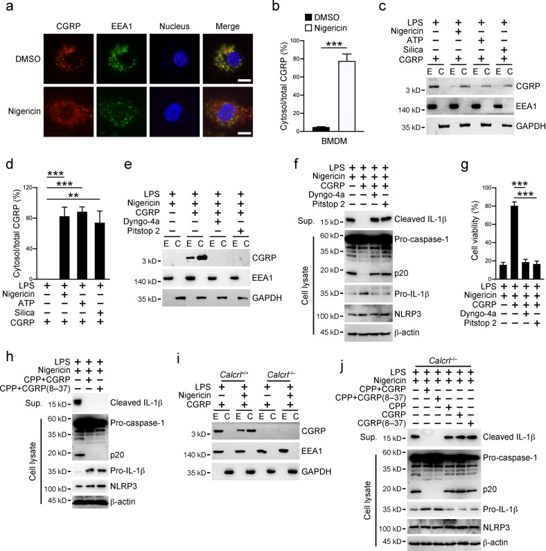
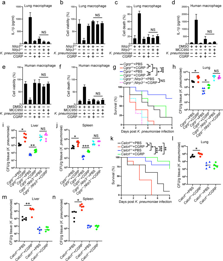
The NLRP3 inflammasome plays an essential role in resistance to bacterial infection. The nervous system secretes multiple neuropeptides affecting the nervous system as well as immune cells. The precise impact of the neuropeptide CGRP on NLRP3 inflammasome activation is still unclear. Here, we show that CGRP negatively regulates the antibacterial process of host cells. CGRP prevents NLRP3 inflammasome activation and reduces mature IL-1β secretion. Following NLRP3 inflammasome stimulation that triggers endosome leakage, CGRP internalized to endosomal compartments is released into the cell cytosol. Cytosolic CGRP binds directly to NLRP3 and dismantles the NLRP3-NEK7 complex, which is crucial for NLRP3 inflammasome activation. CGRP administration exacerbates bacterial infection, while the treatment with a CGRP antagonist has the opposite effect. Our study uncovers a unique role of CGRP in inhibiting inflammasome activation during infections, which might shed new light on antibacterial therapies in the future.

Keywords: CGRP; Inflammasome; NLRP3; Neuropeptide; Suppressor.


Conflict of interest statement

The authors declare no competing interests.

Figures

Fig. 1
Fig. 1
CGRP suppresses host immunity against bacterial infection. a Cgrp+/+ and Cgrp–/– mice were inoculated intratracheally with 3 × 104 CFU of K. pneumoniae, followed by survival rate examinations within a period of 8 days. n = 10 for each group. bd Cgrp+/+ and Cgrp–/– mice were inoculated intratracheally with 3 × 104 CFU of K. pneumoniae for 2 days, followed by bacterial load determinations using homogenates from the lung (b), liver (c) or spleen (d). n = 5 for each group. e Lung pathologies were examined in lungs from the Cgrp+/+ and Cgrp–/– mice inoculated intratracheally with 3 × 104 CFU of K. pneumoniae for 2 days. Scale bar, 50 μm. f Cgrp+/+ and Cgrp–/– mice were inoculated intratracheally with 3 × 104 CFU of K. pneumoniae for 24 h, followed by ELISAs of the indicated cytokines using supernatants of lung homogenates. g, h Cgrp+/+ and Cgrp–/– mice were inoculated intratracheally with 3 × 104 CFU of K. pneumoniae for 24 h. Bronchoalveolar lavage fluids were collected and subjected to an ELISA for IL-1β protein levels in supernatants (g) or an RT‒PCR analysis for Il1b mRNA levels in the pelleted macrophages (h). i Calcrlflox/flox (Calcrl+/+) and Calcrlflox/flox;Lyz2-Cre (Calcrl–/–) mice were inoculated intratracheally with 3 × 104 CFU of K. pneumoniae, followed by survival rate examinations within a period of 8 days. n = 10 for each group. jl Calcrlflox/flox (Calcrl+/+) and Calcrlflox/flox;Lyz2-Cre (Calcrl–/–) mice were inoculated intratracheally with 3 × 104 CFU of K. pneumoniae for 2 days, followed by bacterial load determinations using homogenates from lung (j), liver (k) or spleen (l). n = 5 for each group. m Calcrlflox/flox (Calcrl+/+) and Calcrlflox/flox;Lyz2-Cre (Calcrl–/–) mice were inoculated intratracheally with 3 × 104 CFU of K. pneumoniae for 24 h. Bronchoalveolar lavage fluids were collected and subjected to an ELISA for IL-1β protein levels in supernatants. p.i. post infection, NS nonsignificant. Data are shown as the means ± SDs. *P < 0.05; **P < 0.01; ***P < 0.001. Experiments were repeated three times with similar results
Fig. 2
Fig. 2
CGRP specifically inhibits the NLRP3 inflammasome. ac WT BMDMs primed with 1 μg/ml LPS for 3 h were stimulated with 10 μM nigericin for 30 min, 2 mM ATP for 30 min, 0.5 μM gramicidin for 1 h, 2 μg/ml poly(dA:dT) for 4 h or Salmonella at an MOI of 10 for 4 h with or without the presence of 25 ng/ml CGRP. Cell culture supernatants and cell pellets were collected for immunoblotting with antibodies against the indicated proteins (a). Percentages of viable cells were determined by examining cellular ATP levels in cell pellets (b), and percentages of cells undergoing pyroptosis were calculated by assessing released LDH in cell culture supernatants (c). df WT BMDMs primed with 1 μg/ml LPS for 3 h were stimulated with 2 μM LLOMe, 500 μg/ml silica, 250 μg/ml Alum, 200 μg/ml MSU, 200 μg/ml CPPD, and 200 μg/ml Nano-SiO2 for 6 h with or without the presence of 25 ng/ml CGRP. Cell culture supernatants and cell pellets were collected for immunoblotting with antibodies against the indicated proteins (d). Percentages of viable cells were determined by examining cellular ATP levels in cell pellets (e), and percentages of cells undergoing pyroptosis were calculated by assessing released LDH in cell culture supernatants (f). g Calcrl+/+ and Calcrl–/– BMDMs primed with 1 μg/ml LPS for 3 h were stimulated with 10 μM nigericin for 30 min, 2 mM ATP for 30 min or 500 μg/ml silica for 6 h in the presence of 25 ng/ml CGRP. Cell culture supernatants and cell pellets were collected for immunoblotting with antibodies against the indicated proteins. h WT BMDMs primed with 1 μg/ml LPS for 3 h were stimulated with 10 μM nigericin for 30 min, 2 mM ATP for 30 min or 500 μg/ml silica for 6 h in the presence of 25 ng/ml CGRP or CGRP (8–37). Cell culture supernatants and cell pellets were collected for immunoblotting with antibodies against the indicated proteins. i Macrophages isolated from mouse lungs were primed with 1 μg/ml LPS for 3 h, followed by incubation with 10 μM nigericin in the presence of 25 ng/ml CGRP or CGRP (8–37) for 30 min. Cell culture supernatants were subjected to ELISA to determine IL-1β protein levels. Sup., supernatants. Data are shown as the means ± SDs. Similar results were observed for at least three repeats
Fig. 3
Fig. 3
CGRP enters macrophage cytosol. a, b WT BMDMs primed with 1 μg/ml LPS for 3 h were further incubated with 25 ng/ml CGRP with or without 10 μM nigericin for 30 min. Cells were fixed and stained with antibodies against CGRP and EEA1 (a). Percentages of cytosolic CGRP versus total CGRP inside one cell were calculated, and at least 100 cells were counted (b). Scale bar, 10 μm. c, d WT BMDMs primed with 1 μg/ml LPS for 3 h were stimulated with 10 μM nigericin for 30 min, 2 mM ATP for 30 min or 500 μg/ml silica for 6 h in the presence of 25 ng/ml CGRP. Cell pellets were then subjected to endosome isolation and cell fractionation, followed by immunoblotting with antibodies against the indicated proteins (c). Percentages of cytosolic CGRP versus total CGRP inside cells were calculated (d). e WT BMDMs primed with 1 μg/ml LPS for 3 h were stimulated with 10 μM nigericin together with 25 ng/ml CGRP for 30 min in the presence of 20 μM Pitstop 2 or 10 nM Dyngo-4a as indicated. Cell pellets were then subjected to endosome isolation and cell fractionation, followed by immunoblotting with antibodies against the indicated proteins. f, g WT BMDMs primed with 1 μg/ml LPS for 3 h were stimulated with 10 μM nigericin together with 25 ng/ml CGRP for 30 min in the presence of 20 μM Pitstop 2 or 10 nM Dyngo-4a. Cell culture supernatants and cell pellets were collected for immunoblotting with antibodies against the indicated proteins (f). The percentages of viable cells were determined by examining cellular ATP levels in cell pellets (g). h WT BMDMs were transfected with peptides CGRP or CGRP (8–37) through cell-penetrating peptides (CPP) for 3 h. Cells were then primed with 1 μg/ml LPS for 3 h and stimulated with 10 μM nigericin for 30 min. Cell culture supernatants and cell pellets were collected for immunoblotting with antibodies against the indicated proteins. i Calcrl+/+ and Calcrl–/– BMDMs primed with 1 μg/ml LPS for 3 h were incubated with 25 ng/ml CGRP with or without 10 μM nigericin for 30 min. Cell pellets were then subjected to endosome isolation and cell fractionation, followed by immunoblotting with antibodies against the indicated proteins. j Calcrl–/– BMDM cells were transfected with the peptides CGRP or CGRP (8–37) through cell-penetrating peptides (CPP) for 3 h. Cells were then primed with 1 μg/ml LPS for 3 h and stimulated with 10 μM nigericin for 30 min. Otherwise, Calcrl–/– BMDM cells primed with 1 μg/ml LPS for 3 h were stimulated with 10 μM nigericin together with 25 ng/ml CGRP or CGRP (8–37) for 30 min. Cell culture supernatants and cell pellets were collected for immunoblotting with antibodies against the indicated proteins. E endosome, C cytosol, Sup. supernatants. Data are shown as the means ± SDs. ***P < 0.001. Experiments were repeated three times with similar results
Fig. 4
Fig. 4
CGRP interacts with NLRP3. a Plasmids encoding Gal4 DNA binding domain (BD)-tagged CGRP and Gal4 activating domain (AD)-tagged NLRP3 were cotransfected into the yeast strain AH109. Transformants were grown in the indicated selection media. b Plasmids encoding FLAG-tagged NLRP3, Myc-tagged GFP or GFP-CGRP were cotransfected into HEK293T cells for 24 h, followed by immunoprecipitation with antibody against Myc or with control IgG. Immunoprecipitates and cell lysates were immunoblotted with antibodies against the indicated proteins. c Recombinant GST or GST-tagged CGRP was incubated with cell lysates of HEK293T cells overexpressing FLAG-tagged NLRP3, followed by a GST pulldown assay. Precipitates and cell lysates were immunoblotted with antibodies against the indicated proteins. d WT BMDMs primed with 1 μg/ml LPS for 3 h were stimulated with 10 μM nigericin with or without 25 ng/ml CGRP for 30 min. Cells were then lysed and subjected to immunoprecipitation with an antibody against NLRP3 or control IgG. Immunoprecipitates and cell lysates were immunoblotted with antibodies against the indicated proteins. e Schemes for mouse NLRP3 truncations. f Plasmids encoding FLAG-tagged NLRP3 N-terminal truncations, Myc-tagged GFP or GFP-CGRP were cotransfected into HEK293T cells for 24 h, followed by immunoprecipitation with antibody against FLAG or with control IgG. Immunoprecipitates and cell lysates were immunoblotted with antibodies against the indicated proteins. g Plasmids encoding FLAG-tagged NLRP3 C-terminal truncations, Myc-tagged GFP or GFP-CGRP were cotransfected into HEK293T cells for 24 h, followed by immunoprecipitation with antibody against FLAG or with control IgG. Immunoprecipitates and cell lysates were immunoblotted with antibodies against the indicated proteins. h Nlrp3–/– iBMDMs restored with FLAG-tagged full-length or truncated isoforms of NLRP3 were primed with 1 μg/ml LPS for 3 h and then stimulated with 10 μM nigericin together with 25 ng/ml CGRP for 30 min. Cells were then lysed and subjected to immunoprecipitation with an antibody against FLAG or with control IgG. Immunoprecipitates and cell lysates were immunoblotted with antibodies against the indicated proteins. i, j Nlrp3–/– iBMDMs restored with full-length or truncated isoforms of NLRP3 were primed with 1 μg/ml LPS for 3 h and then stimulated with 10 μM nigericin for 30 min. Cell culture supernatants were collected for ELISAs to determine the secreted IL-1β protein levels (i). The percentages of viable cells were determined by examining cellular ATP levels in cell pellets (j). Data are shown as the means ± SDs. Similar results were observed for at least three repeats
Fig. 5
Fig. 5
CGRP expels NEK7 from NLRP3. a FLAG-tagged NLRP3 was immunoprecipitated from HEK293T cells overexpressing FLAG-NLRP3 through anti-FLAG antibody-conjugated beads. Beads containing NLRP3 were incubated with 100 μCi [γ-32P]ATP in the presence of 100 ng/ml CGRP or 10 mM ATP, followed by washing with PBS every 10 min. The signals of radioactive ATP were detected through liquid scintillation. b Plasmids encoding FLAG-tagged NLRP3, HA-tagged NEK7 and increasing amounts of Myc-tagged GFP or GFP-CGRP were cotransfected into HEK293T cells for 24 h, followed by immunoprecipitation with antibody against FLAG or with control IgG. Immunoprecipitates and cell lysates were immunoblotted with antibodies against the indicated proteins. c Plasmids encoding FLAG-tagged NLRP3, Myc-tagged GFP or GFP-CGRP and increasing amounts of HA-tagged NEK7 were cotransfected into HEK293T cells for 24 h, followed by immunoprecipitation with antibody against FLAG or with control IgG. Immunoprecipitates and cell lysates were immunoblotted with antibodies against the indicated proteins. d Plasmids encoding FLAG-tagged NLRP3, Myc-tagged GFP or GFP-CGRP and HA-tagged NEK7 were cotransfected into HEK293T cells for 24 h, followed by immunoprecipitation with an antibody against Myc or with control IgG. Immunoprecipitates and cell lysates were immunoblotted with antibodies against the indicated proteins. e Plasmids encoding Myc-tagged GFP or GFP-CGRP and HA-tagged NEK7 were cotransfected into HEK293T cells for 24 h, followed by immunoprecipitation with antibody against Myc or with control IgG. Immunoprecipitates and cell lysates were immunoblotted with antibodies against the indicated proteins. f Plasmids encoding Myc-tagged GFP or GFP-CGRP, FLAG-tagged NLRP3 and HA-tagged NEK7 were cotransfected into HEK293T cells for 24 h, followed by immunoprecipitation with antibody against HA or with control IgG. Immunoprecipitates and cell lysates were immunoblotted with antibodies against the indicated proteins. g WT BMDMs primed with 1 μg/ml LPS for 3 h were stimulated with 10 μM nigericin with or without 25 ng/ml CGRP for 30 min. Cells were lysed and subjected to immunoprecipitation with an antibody against NLRP3 or with control IgG. Immunoprecipitates and cell lysates were immunoblotted with antibodies against the indicated proteins. h, i WT BMDMs (h) or Calcrl knockout cells (i) primed with 1 μg/ml LPS for 3 h were stimulated with 10 μM nigericin with or without 25 ng/ml CGRP for 30 min. Cells were lysed and subjected to immunoprecipitation with an antibody against NLRP3 or with control IgG. Immunoprecipitates and cell lysates were immunoblotted with antibodies against the indicated proteins. j Nlrp3–/– iBMDMs restored with FLAG-tagged full-length or truncated isoforms of NLRP3 were primed with 1 μg/ml LPS for 3 h and then stimulated with 10 μM nigericin for 30 min. Cells were then lysed and subjected to immunoprecipitation with an antibody against FLAG or with control IgG. Immunoprecipitates and cell lysates were immunoblotted with antibodies against the indicated proteins. k Nlrp3–/– iBMDMs restored with full-length or truncated isoforms of NLRP3 were primed with 1 μg/ml LPS for 3 h and then stimulated with 10 μM nigericin for 30 min. Cells were then fixed and stained with an antibody against ASC, followed by the examination of ASC specks inside cells. One hundred cells were assessed for each group. Data are shown as the means ± SDs. Experiments were repeated three times with similar results
Fig. 6
Fig. 6
CGRP administration exacerbates bacterial infection. ac Macrophages from Nlrp3+/+ and Nlrp3–/– lungs primed with 1 μg/ml LPS for 3 h were incubated with K. pneumoniae at an MOI of 10 for 1 h with or without the presence of 25 ng/ml CGRP. Cell culture supernatants were collected for ELISAs to determine the secreted IL-1β protein levels (a). Percentages of viable cells were determined by examining cellular ATP levels in cell pellets (b), and percentages of cells undergoing pyroptosis were calculated by assessing released LDH in cell culture supernatants (c). df Macrophages derived from human peripheral blood mononuclear cells were primed with 1 μg/ml LPS for 3 h and then incubated with K. pneumoniae at an MOI of 10 for 1 h with or without the presence of 25 ng/ml CGRP. Cell culture supernatants were collected for ELISAs to determine the secreted IL-1β protein levels (d). Percentages of viable cells were determined by examining cellular ATP levels in cell pellets (e), and percentages of cells undergoing pyroptosis were calculated by assessing released LDH in cell culture supernatants (f). g Cgrp+/+, Cgrp–/– and Cgrp–/–;Nlrp3–/–mice were inoculated intratracheally with 3 × 104 CFU of K. pneumoniae. Moreover, 2 μg CGRP was administered intranasally to each mouse once a day. Survival rates were examined within a period of 8 days. n = 10 for each group. hj Cgrp+/+, Cgrp–/– and Cgrp–/–;Nlrp3–/– mice were inoculated intratracheally with 3 × 104 CFU of K. pneumoniae. Moreover, 2 μg CGRP was administered intranasally to each mouse once a day. Bacterial load determinations using homogenates from lung (h), liver (i) or spleen (j) were performed 2 days later. n = 5 for each group. k Calcrlflox/flox (Calcrl+/+) and Calcrlflox/flox;Lyz2-Cre (Calcrl–/–) mice were inoculated intratracheally with 3 × 104 CFU of K. pneumoniae. Moreover, 2 μg CGRP was administered intranasally to each mouse once a day. Survival rates were examined within a period of 8 days. n = 10 for each group. ln Calcrlflox/flox (Calcrl+/+) and Calcrlflox/flox;Lyz2-Cre (Calcrl–/–) mice were inoculated intratracheally with 3 × 104 CFU of K. pneumoniae. Moreover, 2 μg CGRP was administered intranasally to each mouse once a day. Bacterial load determinations using homogenates from lung (l), liver (m) or spleen (n) were performed 2 days later. n = 5 for each group. Data are shown as the means ± SDs. *P < 0.05; **P < 0.01; ***P < 0.001. Similar results were observed for at least three repeats

References

    1. Fitzgerald KA, Kagan JC. Toll-like receptors and the control of immunity. Cell. 2020;180:1044–66. doi: 10.1016/j.cell.2020.02.041. - DOI - PMC - PubMed
    1. Liu G, Gack MU. Distinct and orchestrated functions of RNA sensors in innate immunity. Immunity. 2020;53:26–42. doi: 10.1016/j.immuni.2020.03.017. - DOI - PMC - PubMed
    1. Charmoy M, Hurrell BP, Romano A, Lee SH, Ribeiro-Gomes F, Riteau N, et al. The Nlrp3 inflammasome, IL-1β, and neutrophil recruitment are required for susceptibility to a nonhealing strain of Leishmania major in C57BL/6 mice. Eur J Immunol. 2016;46:897–911. doi: 10.1002/eji.201546015. - DOI - PMC - PubMed
    1. Mitchell PS, Sandstrom A, Vance RE. The NLRP1 inflammasome: new mechanistic insights and unresolved mysteries. Curr Opin Immunol. 2019;60:37–45. doi: 10.1016/j.coi.2019.04.015. - DOI - PMC - PubMed
    1. Sharma BR, Kanneganti TD. NLRP3 inflammasome in cancer and metabolic diseases. Nat Immunol. 2021;22:550–9. doi: 10.1038/s41590-021-00886-5. - DOI - PMC - PubMed

Publication types

MeSH terms

Substances