. 2023 Jan 4;49(1):2.
doi: 10.1186/s13052-022-01401-8.

SIGIRR-caspase-8 signaling mediates endothelial apoptosis in Kawasaki disease

Affiliations

SIGIRR-caspase-8 signaling mediates endothelial apoptosis in Kawasaki disease

Zhengwang Wen et al. Ital J Pediatr. .

Abstract

Background: Kawasaki disease (KD) is a kind of vasculitis with unidentified etiology. Given that the current diagnosis and therapeutic strategy of KD are mainly dependent on clinical experiences, further research to explore its pathological mechanisms is warranted.

Methods: Enzyme linked immunosorbent assay (ELISA) was used to measure the serum levels of SIGIRR, TLR4 and caspase-8. Western blotting was applied to determine protein levels, and flow cytometry was utilized to analyze cell apoptosis. Hematoxylin eosin (HE) staining and TUNEL staining were respectively used to observe coronary artery inflammation and DNA fragmentation.

Results: In this study, we found the level of SIGIRR was downregulated in KD serum and KD serum-treated endothelial cells. However, the level of caspase-8 was increased in serum from KD patients compared with healthy control (HC). Therefore, we hypothesized that SIGIRR-caspase-8 signaling may play an essential role in KD pathophysiology. In vitro experiments demonstrated that endothelial cell apoptosis in the setting of KD was associated with caspase-8 activation, and SIGIRR overexpression alleviated endothelial cell apoptosis via inhibiting caspase-8 activation. These findings were also recapitulated in the Candida albicans cell wall extracts (CAWS)-induced KD mouse model.

Conclusion: Our data suggest that endothelial cell apoptosis mediated by SIGIRR-caspase-8 signaling plays a crucial role in coronary endothelial damage, providing potential targets to treat KD.

Keywords: Apoptosis; Caspase-8; Endothelial cell; Kawasaki disease; SIGIRR.


Conflict of interest statement

None.

Figures

Fig. 1
Fig. 1
Levels of SIGIRR, TLR4 and/or caspase-8 in KD serum and KD serum-treated endothelial cells were elevated. A-C The levels of SIGIRR, TLR4 and caspase-8 were measured in the sera from HC, Febrile, Acute KD and Conv KD by ELISA (n = 11). D-F The protein expression of SIGIRR and TLR4 were determined in the treated endothelial cells by Western blot analysis. Data were shown as mean ± SD (n = 3). * P < 0.05 indicated the significant difference
Fig. 2
Fig. 2
Endothelial cell apoptosis in KD condition was associated with caspase-8 activation. A The apoptotic percentage of endothelial cells was analyzed using flow cytometry. The histogram was the quantitative analysis of Annexin V-FITC-positive cells. B-G The protein expression levels of SIGIRR, TLR4, cleaved caspase-8 and cleaved caspase-3, and BAX/Bcl-2 ratio were determined in the treated endothelial cells by Western blotting. Data were presented as mean ± SD (n = 3). * P < 0.05 versus HC group, and # P < 0.05 versus KD group
Fig. 3
Fig. 3
SIGIRR overexpression rescued endothelial cell apoptosis. A-F The protein expression levels of SIGIRR, TLR4, cleaved caspase-8 and cleaved caspase-3, and BAX/Bcl-2 ratio were determined in the treated endothelial cells by Western blotting. G The apoptotic percentage of endothelial cells was analyzed using flow cytometry. The histogram was the quantitative analysis of Annexin V-FITC-positive cells. Data were expressed as mean ± SD (n = 3).* P < 0.05 versus HC group, and # P < 0.05 versus KD group
Fig. 4
Fig. 4
SIGIRR mitigated endothelial cell apoptosis via inhibiting caspase-8 activity. A The apoptotic percentage of endothelial cells was analyzed using flow cytometry. B-G The protein expression levels of SIGIRR, TLR4, cleaved caspase-8 and cleaved caspase-3, and BAX/Bcl-2 ratio were determined in the treated endothelial cells by Western blotting. * P < 0.05 versus HC group, # P < 0.05 versus KD group, and & P < 0.05 versus KD + AAV-SIGIRR group
Fig. 5
Fig. 5
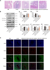
SIGIRR overexpression alleviated coronary artery injury in a murine model of KD. A Heart tissues were harvested on 28 day after CAWS injection, and the representative images from H&E staining were shown. Magnification: × 200. Scale bar = 100 μm. B-G The protein expression of SIGIRR, TLR4, cleaved caspase-8 and cleaved caspase-3, and BAX/Bcl-2 ratio in the heart tissues were determined by Western blotting. H DNA fragmentation in the endothelial cells was assessed by co-localized staining of TUNEL (green) and CD31 (an endothelial marker, red) (n = 5). Magnification: × 200. Scale bar = 50 μm. * P < 0.05 versus PBS group, and # P < 0.05 versus CAWS group
Fig. 6
Fig. 6
SIGIRR relieved coronary artery injury via inhibiting caspase-8 activation. A The representative images from H&E staining were shown. Magnification: × 200. Scale bar = 100 μm. B-G The protein expression of SIGIRR, TLR4, cleaved caspase-8 and cleaved caspase-3, and BAX/Bcl-2 ratio in the heart tissues were detected by Western blotting. H DNA fragmentation in the coronary endothelium was evaluated by co-localized staining of TUNEL (green) and CD31 (an endothelial marker, red) (n = 5). Magnification: × 200. Scale bar = 50 μm. * P < 0.05 versus PBS group, # P < 0.05 versus CAWS group, and & P < 0.05 versus CAWS + AAV-SIGIRR group

Similar articles

Cited by

References

    1. Rife E, Gedalia A. Kawasaki disease: an update. Curr Rheumatol Rep. 2020;22:1–10. doi: 10.1007/s11926-020-00941-4. - DOI - PMC - PubMed
    1. Newburger JW, Takahashi M, Burns JC. Kawasaki disease. J Am Coll Cardiol. 2016;67:1738–1749. doi: 10.1016/j.jacc.2015.12.073. - DOI - PubMed
    1. He M, Chen Z, Martin M, Zhang J, Sangwung P, Woo B, Tremoulet AH, Shimizu C, Jain MK, Burns JC, Shyy JY. MiR-483 Targeting of CTGF Suppresses Endothelial-to-Mesenchymal Transition: Therapeutic Implications in Kawasaki Disease. Circ Res. 2017;120:354–365. doi: 10.1161/CIRCRESAHA.116.310233. - DOI - PMC - PubMed
    1. Chu M, Wu R, Qin S, Hua W, Shan Z, Rong X, Zeng J, Hong L, Sun Y, Liu Y, Li W, Wang S, Zhang C. Bone Marrow-Derived MicroRNA-223 Works as an Endocrine Genetic Signal in Vascular Endothelial Cells and Participates in Vascular Injury From Kawasaki Disease. J Am Heart Assoc. 2017;6:1–14. doi: 10.1161/JAHA.116.004878. - DOI - PMC - PubMed
    1. Jia C, Zhuge Y, Zhang S, Ni C, Wang L, Wu R, Niu C, Wen Z, Rong X, Qiu H, Chu M. IL-37b alleviates endothelial cell apoptosis and inflammation in Kawasaki disease through IL-1R8 pathway. Cell Death Dis. 2021;12:575. doi: 10.1038/s41419-021-03852-z. - DOI - PMC - PubMed