. 2022 Dec;10(12):e005669.
doi: 10.1136/jitc-2022-005669.

Systemic cellular viroimmunotherapy for canine high-grade gliomas

Affiliations

Systemic cellular viroimmunotherapy for canine high-grade gliomas

Ana Cloquell et al. J Immunother Cancer. 2022 Dec.

Abstract

Background: Oncolytic viruses constitute a growing field of interest, both in human and veterinary oncology, given that they are particularly helpful for treating non-surgical tumors and disseminated cancer, such as high-grade gliomas. Companion dogs present malignant gliomas with biological, genetic, phenotypic, immunological, and clinical similarities to human gliomas. These features favor comparative approaches, leading to the treatment of canine oncological patients to achieve translational applications to the human clinic. The systemic administration of oncolytic viruses presents a challenge due to their limitations in effectively targeting tumors and metastases. Therefore, the aim of this study is to evaluate the safety and antitumor activity of a virotherapy used in spontaneous canine tumors.

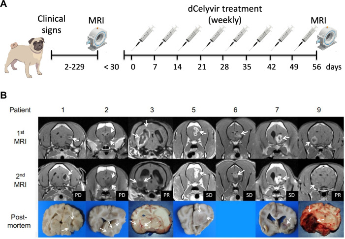
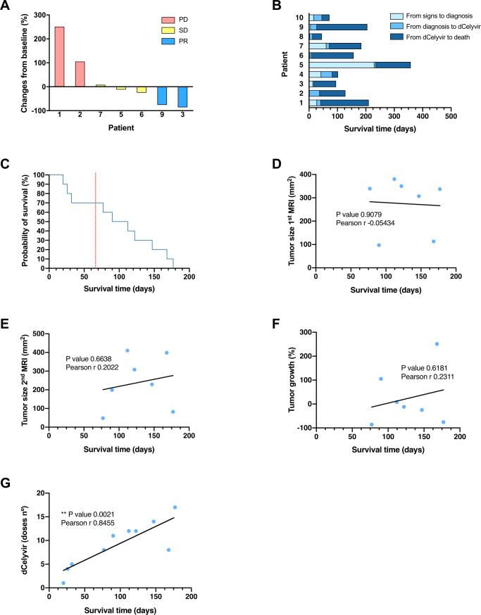
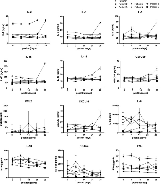
Methods: Ten dogs with high-grade rostrotentorial gliomas underwent weekly systemic endovenous cellular virotherapy with dCelyvir (canine mesenchymal stem cells infected with the canine oncolytic adenovirus ICOCAV17) for 8 weeks. Efficacy was determined in seven dogs according to the Response Assessment in Veterinary Neuro-Oncology criteria considering clinical status and MRI measurements. Medical history, physical and neurological examinations, and vaccination status were evaluated prior to and during follow-up. Safety was evaluated by physical examinations and hematological and biochemical changes in peripheral blood. Immune populations were analyzed by flow cytometry in peripheral blood and by gene expression and immunohistochemistry in the tumor microenvironment.

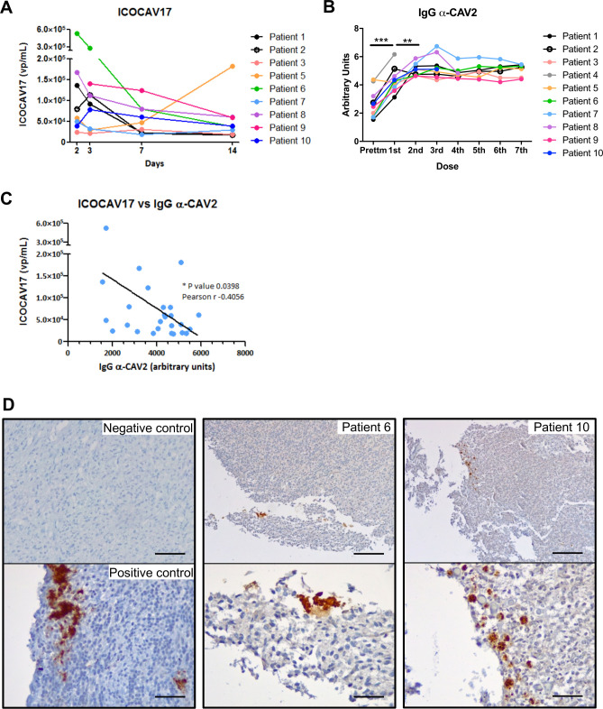
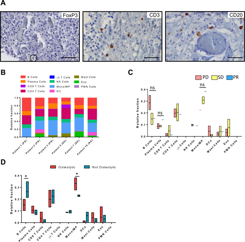
Results: The treatment was well tolerated and major adverse effects were not observed. Two dogs had partial responses (76% and 86% reduction in tumor size), and 3/7 showed stable disease. ICOCAV17 was detected in peripheral blood in nine dogs, and a correlation between the ICOCAV17 particles and anti-canine adenovirus (CAV) antibodies was observed. ICOCAV17 was detected in 3/9 tumor tissues after necropsies. Regarding tumor-infiltrating lymphocytes, the dogs with disease stabilization and partial response tended to have reduced memory B-cell infiltration and increased monocyte/macrophage lineage cells.

Conclusions: These findings indicate that dCelyvir is safe and presents efficacy in canine rostrotentorial high-grade gliomas. These data are relevant to the ongoing phase Ib regulated human clinical trial that is administering this virotherapy to children, adolescents, and young adults with diffuse pontine glioma. Celyvir should be further explored as a treatment in veterinary and human neuro-oncology.

Keywords: Brain Neoplasms; Central Nervous System Neoplasms; Immunotherapy; Oncolytic Virotherapy; Oncolytic Viruses.


Conflict of interest statement

Competing interests: None declared.

Figures

Figure 1
Figure 1
(A)Schematic representation of the dCelyvir treatment. (B) MRI and macroscopic tumor images from the dCelyvir-treated patients. MRI prior to dCelyvir treatment (first MRI), and 2 months after starting weekly treatment (second MRI) showing tumors (white arrows). Clinical benefit is indicated according to RAVNO criteria. Bottom figures show macroscopic tumor tissue (white arrows) in necropsies. PD, progressive disease; PR, partial response; RAVNO, response assessment in veterinary neuro-oncology; SD, stable disease.
Figure 2
Figure 2
Tumor growth and survival time from the dCelyvir-treated patients. (A) Waterfall plot showing changes in tumor size. Tumor growth from baseline (first MRI) to 2 months after starting the dCelyvir treatment (second MRI) is shown for each patient. (B) Survival time. Bar plot showing the total survival time for each patient. The days from symptom onset to diagnosis, from diagnosis to starting dCelyvir treatment, and from treatment to death are indicated for each patient. (C) Survival analysis. Kaplan-Meier curve showing the probability of survival for dCelyvir-treated dogs (blue line) starting with day of treatment up to death. The dashed red line indicates the MST according to the literature and the historical cases treated with resective surgery. (D–G) Correlation analysis between survival time and tumor size at first MRI (D), at second MRI (E), tumor growth (F), and dCelyvir dose (G) calculated by Pearson’s correlation coefficient (r). Individual values (blue dots) and regression lines are shown. **p<0.005. MST, median survival time; PD, progressive disease; PR, partial response; SD, stable disease.
Figure 3
Figure 3
Oncolytic virus and antibody response. (A) Adenovirus detection during follow-up. Adenovirus presence analyzed by quantitative PCR (qPCR) in peripheral blood is shown. Quantification time refers to the time after the first day of treatment (day 0). Individual values from each patient are shown. (B)Specific antibodies. IgG α-CAV2 quantified by solid phase ELISA in the serum of canine patients during dCelyvir treatments. A Wilcoxon test was performed (***p<0.0001; **p<0.005). (C)Correlation between oncolytic adenovirus and specific antibodies was calculated by Pearson’s correlation coefficient (r), using data from all available dogs up to the second week of treatment (14th day after treatment). Individual values (blue dots) and a regression line are shown. *p<0.05. (D)The presence of adenovirus inside gliomas analyzed by immunohistochemistry. Representative images showing adenovirus-positive cells (brown) on formalin-fixed, paraffin-embedded-positive control tissue, and tumor necropsies from patients 6 and 10. Scale bars: 50 µm (negative and positive controls), 200 µm (top patients) and 50 µm (bottom patients).
Figure 4
Figure 4
Cytokines in peripheral blood from treated patients quantified by a multiplex assay. Sera from treated dogs were analyzed prior to treatment (d0) and Weekly after the first four treatments (days 7, 14, 21, and 28) for IL-2, IL-6, IL-7, IL-15, IL-18, GM-CSF, CCL2, CXCL10, IL-8, IL-10, KC-like, and IFN-ɣ. Individual values are shown. GM-CSF, granulocyte-macrophage colony-stimulating factor.
Figure 5
Figure 5
Intratumor immune microenvironment characterization after dCelyvir treatment. (A) The presence of immune cells inside gliomas analyzed by immunohistochemistry. Representative images showing immune cells (brown) on formalin-fixed, paraffin-embedded tumors at necropsies. Scale bars: 50 µm. (B) Relative fraction of 11 types of TILs in 6 dogs estimated with CIBERSORT. (C) Immune cell relative fraction from patients depending on their clinical benefit, after dCelyvir treatment. (D) Immune cell relative fraction from patients, comparing those that present an osteolytic pattern at CT imaging (osteolytic) and those that do not (not osteolytic). Statistic test: Two-way ANOVA and Bonferroni’s multiple comparison test; Alpha: 0.05; ns: not significant; *p<0.05. ANOVA, analysis of variance; DC, dendritic cells; Eos, Eosinophils; Mono/MP, Monocyte/Macrophages; PD, progressive disease; PMN, polymorphonuclear cells; PR, partial response; SD, stable disease; TILs, tumor-infiltrating lymphocytes.

References

    1. Sánchez D, Cesarman-Maus G, Amador-Molina A, et al. Oncolytic viruses for canine cancer treatment. Cancers 2018;10:404. 10.3390/cancers10110404 - DOI - PMC - PubMed
    1. Russell L, Peng KW, Russell SJ, et al. Oncolytic viruses: priming time for cancer immunotherapy. BioDrugs 2019;33:485–501. 10.1007/s40259-019-00367-0 - DOI - PMC - PubMed
    1. Tähtinen S, Grönberg-Vähä-Koskela S, Lumen D, et al. Adenovirus improves the efficacy of adoptive T-cell therapy by recruiting immune cells to and promoting their activity at the tumor. Cancer Immunol Res 2015;3:915–25. 10.1158/2326-6066.CIR-14-0220-T - DOI - PubMed
    1. Woller N, Gürlevik E, Fleischmann-Mundt B, et al. Viral infection of tumors overcomes resistance to PD-1-immunotherapy by broadening Neoantigenome-directed T-cell responses. Mol Ther 2015;23:1630–40. 10.1038/mt.2015.115 - DOI - PMC - PubMed
    1. Raja J, Ludwig JM, Gettinger SN, et al. Oncolytic virus immunotherapy: future prospects for oncology. J Immunother Cancer 2018;6:140. 10.1186/s40425-018-0458-z - DOI - PMC - PubMed

Publication types