

Computed Tomography Scan Architectural Measurements in Adult Foot and Ankle Surgery: A Narrative Review for Orthopaedic Trainees
- PMID: 36600866
- PMCID: PMC9801486
- DOI: 10.7759/cureus.32039
Computed Tomography Scan Architectural Measurements in Adult Foot and Ankle Surgery: A Narrative Review for Orthopaedic Trainees
Abstract
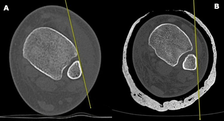
CT scan plays an important role in adult foot and ankle surgery. Plain radiographs are usually the first-line imaging modality for assessing foot and ankle bone and joint architectural abnormalities. However, despite the fact that a CT scan is more expensive and associated with higher radiation exposure, it offers better imaging quality for the assessment of bony lesions in orthopaedics and trauma. Evidence has shown that more accurate measurements can be obtained from a CT scan compared to plain radiographs. Weight-bearing multi-detection CT scanning goes the extra mile by providing a more detailed assessment, especially for intra-articular fractures, and mirrors the real-life foot and ankle dynamics compared to conventional non-weight-bearing CT scans. It also has a relatively lower radiation dose compared to conventional CT scans. CT scan is the best modality for assessing bony lesions whereas MRI is better for soft tissue pathology. An understanding of the role of CT scan in the anatomical assessment of the foot and ankle will help improve communication between orthopaedic surgeons, radiologists, and radiographers. A thorough understanding of when to use a CT scan compared to the other imaging modalities will also lead to better surgical outcomes, reduced cost, and reduced risk from radiation exposure. This review article analyzes the role of CT in assessing relevant radiographic architectural measurements for diagnosis and surgical planning in adult foot and ankle surgery.
Keywords: computed tomography scan; ct scan; foot and ankle surgery; foot deformity; radiological findings; weight-bearing ct.
Copyright © 2022, Anazor et al.
Conflict of interest statement
The authors have declared that no competing interests exist.
Figures


References
-
- Weight-bearing CT in foot and ankle pathology. Lintz F, Beaudet P, Richardi G, Brilhault J. Orthop Traumatol Surg Res. 2021;107:102772. - PubMed
-
- Techniques for 3D foot bone orientation angles in weight-bearing from cone-beam computed tomography. Carrara C, Belvedere C, Caravaggi P, Durante S, Leardini A. Foot Ankle Surg. 2021;27:168–174. - PubMed
-
- Feger Feger, J J. Ankle protocol (CT). Reference article. Radiopaedia. [ Nov; 2021 ]. 2021. https://radiopaedia.org/articles/ct-ankle-protocol-1 https://radiopaedia.org/articles/ct-ankle-protocol-1
-
- Feger Feger, J J. Foot protocol (CT). Reference article. Radiopaedia. [ Nov; 2021 ]. 2021. https://radiopaedia.org/articles/ct-foot-protocol-1 https://radiopaedia.org/articles/ct-foot-protocol-1
-
- AO trauma: AO Foundation surgery reference. [ Oct; 2022 ]. 2006. https://surgeryreference.aofoundation.org https://surgeryreference.aofoundation.org
Publication types
LinkOut - more resources
Full Text Sources