. 2022 Nov 30:2022:8287633.
doi: 10.1155/2022/8287633. eCollection 2022.

Suppression of Selective Voltage-Gated Calcium Channels Alleviates Neuronal Degeneration and Dysfunction through Glutathione S-Transferase-Mediated Oxidative Stress Resistance in a Caenorhabditis elegans Model of Alzheimer's Disease

Affiliations

Suppression of Selective Voltage-Gated Calcium Channels Alleviates Neuronal Degeneration and Dysfunction through Glutathione S-Transferase-Mediated Oxidative Stress Resistance in a Caenorhabditis elegans Model of Alzheimer's Disease

Zihui Zheng et al. Oxid Med Cell Longev. .

Abstract

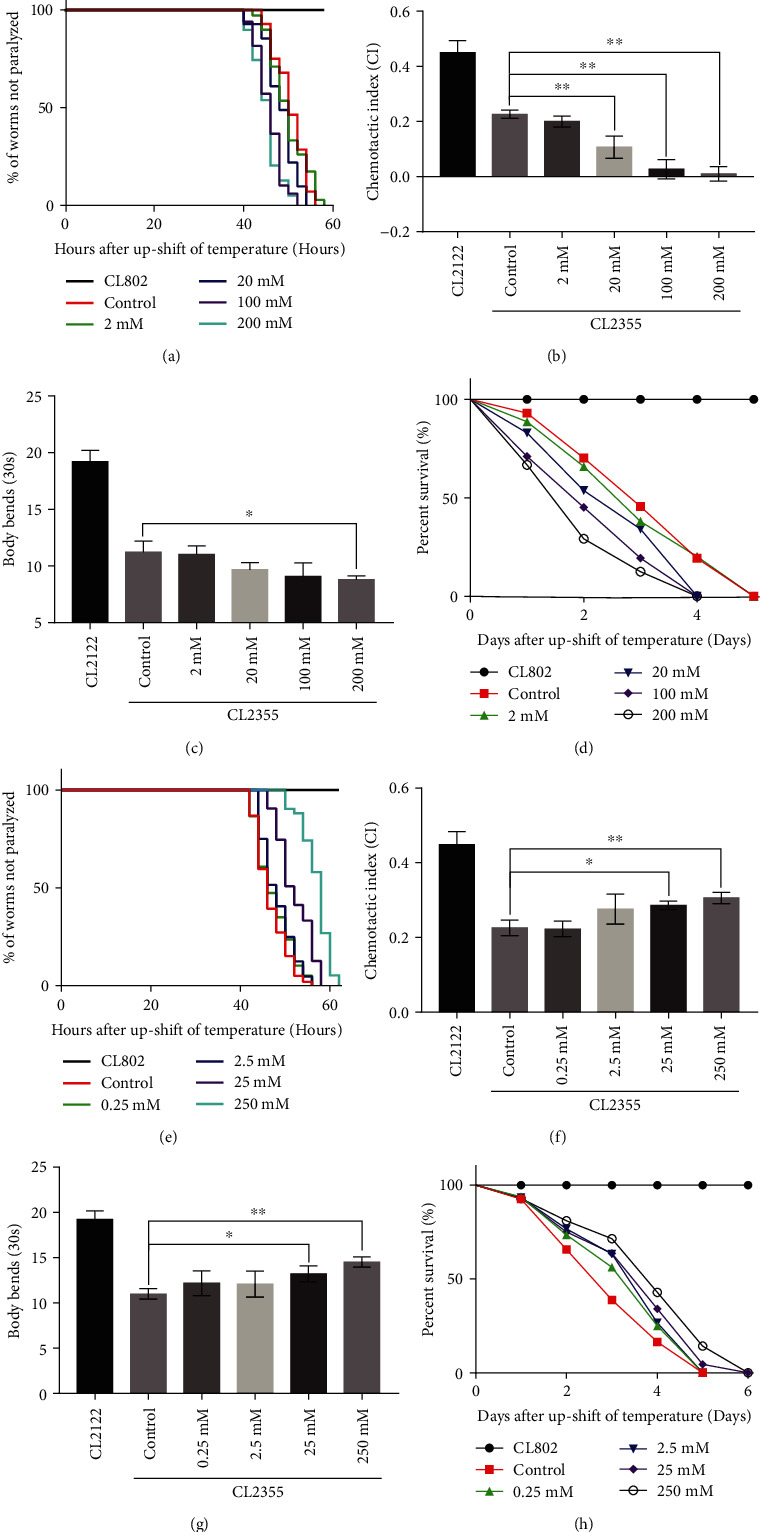
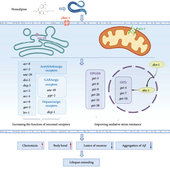
Calcium homeostasis plays a vital role in protecting against Alzheimer's disease (AD). In this study, amyloid-β (Aβ)-induced C. elegans models of AD were used to elucidate the mechanisms underlying calcium homeostasis in AD. Calcium acetate increased the intracellular calcium content, exacerbated Aβ 1-42 aggregation, which is closely associated with oxidative stress, aggravated neuronal degeneration and dysfunction, and shortened the lifespan of the C. elegans models. Ethylene glycol tetraacetic acid (EGTA) and nimodipine were used to decrease the intracellular calcium content. Both EGTA and nimodipine showed remarkable inhibitory effects on Aβ 1-42 aggregations by increasing oxidative stress resistance. Moreover, both compounds significantly delayed the onset of Aβ-induced paralysis, rescued memory deficits, ameliorated behavioral dysfunction, decreased the vulnerability of two major (GABAergic and dopaminergic) neurons and synapses, and extended the lifespan of the C. elegans AD models. Furthermore, RNA sequencing of nimodipine-treated worms revealed numerous downstream differentially expressed genes related to calcium signaling. Nimodipine-induced inhibition of selective voltage-gated calcium channels was shown to activate other calcium channels of the plasma membrane (clhm-1) and endoplasmic reticulum (unc-68), in addition to sodium-calcium exchanger channels (ncx-1). These channels collaborated to activate downstream events to resist oxidative stress through glutathione S-transferase activity mediated by HPGD and skn-1, as verified by RNA interference. These results may be applied for the treatment of Alzheimer's disease.


Conflict of interest statement

The authors declare that this research was conducted with no commercial or financial relationships that could be construed as a potential conflict of interest.

Figures

Figure 1
Figure 1
Calcium acetate aggravated Aβ-induced neurodegenerative motor impairments, while EGTA inhibited neurodegenerative dysfunction and extended the life span of Aβ worms. (a) Time course of Aβ-induced paralysis in CL4176 worms treated with/out calcium acetate. CL802 was the control strain. (b) CI values of calcium acetate -treated vs. untreated CL2355 worms. CL2122 was the control strain. (c) Body bends capacity of calcium acetate-treated vs. untreated CL2355 worms. CL2122 was the control strain. (d) Time course of lifespan of calcium acetate-treated vs. untreated CL4176 worms. CL802 was the control strain. (e) Time course of Aβ-induced paralysis in CL4176 worms treated with/out EGTA. (f) CI values of EGTA-treated vs. untreated CL2355 worms. (g) Body bends capacity of EGTA-treated vs. untreated CL2355 worms. (h) Time course of lifespan of EGTA-treated vs. untreated CL4176 worms. Error bars indicate SEM. (p < 0.05 vs. control, ∗∗p < 0.01 vs. control).
Figure 2
Figure 2
Calcium acetate aggravated impairment of GABAergic and motor neurons in the VNC, while EGTA and nimodipine postponed lesion development in Aβ worms. (a) Representative images of D-type GABAergic neurons in the VNCs of EG1285, NUCM0001, calcium acetate-treated NUCM0001, EGTA-treated NUCM0001, and nimodipine-treated NUCM0001 worms (scale bar, 50 μm). Histogram showing the number of lost D-type GABAergic neurons in NUCM0001 worms treated with/out calcium-adjusting agents. Lost neurons are indicated by triangles. (b) Representative images of motor neurons in the VNCs of BL5717, NUCM0002, calcium acetate -treated NUCM0002, EGTA-treated NUCM0002, and nimodipine-treated NUCM0002 worms (scale bar, 50 μm). Histogram of the fluorescence intensity of cell bodies of NUCM0002 worms treated with/out calcium-adjusting agents. Lost neurons are indicated with triangles. Motor neurons are indicated with arrows. Error bars indicate the SEM. (p < 0.05 vs. control, ∗∗p < 0.01 vs. control).
Figure 3
Figure 3
Calcium acetate aggravated impairment of dopaminergic neurons and synapses, while EGTA and nimodipine postponed the development of Aβ aggregates. (a) Representative images of dopaminergic neurons in UA57, NUCM0003, calcium acetate-treated NUCM0003, EGTA-treated NUCM0003, and nimodipine-treated NUCM0003 worms (scale bar, 50 μm). Histogram of the numbers of lost dopaminergic neurons in NUCM0003 worms treated with/out calcium-adjusting agents. Lost neurons are indicated with triangles. Motor neurons are indicated with arrows. (b) Representative images of presynaptic densities in the VNCs of NC571, NUCM0004, calcium acetate-treated NUCM0004, EGTA-treated NUCM0004, and nimodipine-treated NUCM0004 worms (scale bar, 20 μm). Histogram of the number of gaps in NUCM0004 worms treated with/out calcium-adjusting agents. Gaps are indicated with triangles. Error bars indicate the SEM. (p < 0.05 vs. control, ∗∗p < 0.01 vs. control).
Figure 4
Figure 4
Nimodipine inhibited neuronal degeneration and extended the lifespan of Aβ worms. (a) Time course of Aβ-induced paralysis in CL4176 worms treated with/out nimodipine. CL802 was the control strain. (b) The CI of CL2355 worms treated with/out nimodipine. CL2122 was the control strain. (c) Body bends capacity of CL2355 worms treated with/out nimodipine. CL2122 was the control strain. (d) Time course of the lifespan of CL4176 worms treated with/out nimodipine. CL802 was the control strain. Error bars indicate the SEM. (p < 0.05 vs. control, ∗∗p < 0.01 vs. control).
Figure 5
Figure 5
Calcium acetate aggravated Aβ aggregation by increasing oxidative stress in Aβ worm, while EGTA and nimodipine had opposite effects. (a‑e) Representative images of Aβ deposits in N2, CL2355, and CL2355 worms treated with calcium acetate, EGTA, and nimodipine (scale bar, 20 μm). The area in the red rectangle was observed. Arrows indicate ThS staining. (f) Histogram of the mean number of Aβ deposits in the head region/anterior area of the worms. (g–k) Representative images of ROS content tested by the dihydroethidium (DHE) red fluorescence, in N2, CL2355, and CL2355 worms treated with calcium acetate, EGTA, and nimodipine (scale bar, 100 μm). (l) Histogram of the mean ROS content. Error bars indicate the SEM. (m) Histogram of the mean ATP content in N2, CL2355, and CL2355 worms treated with calcium acetate, EGTA, and nimodipine. (p < 0.05 vs. control, ∗∗p < 0.01 vs. control).
Figure 6
Figure 6
Nimodipine upregulated the transcript levels of genes encoding antioxidative enzymes (HPGDS and GST). (a) Relative mRNA levels of genes relevant to antioxidative stress in CL2355 worms treated with/out nimodipine. (b) Representative images of gst-4P::GFP expression in CL2166 and CL2166 worms treated with PQ, CL2166, and nimodipine (scale bar, 100 μm). (c) Histogram of gst-4P::GFP relative fluorescence intensity/body area of the worm.
Figure 7
Figure 7
Suppression of selective VGCCs enhanced oxidative stress resistance through regulation of HPGDS and GST activities in CL2355 Aβ worms. (a) Activity of clhm-1, unc-68, or ncx-1 in regulating the effect of nimodipine on the CI value. L4440 is an empty vector. (b) Activity of clhm-1, unc-68, or ncx-1 in regulating the effect of nimodipine on body bending capacity. L4440 is an empty vector. (c) Relative mRNA levels of genes relevant to antioxidative stress in CL2355 and CL2355 clhm-1 (RNAi) worms treated with nimodipine (d) Relative mRNA levels of genes relevant to antioxidative stress in CL2355 and CL2355 unc-68 (RNAi) worms treated with nimodipine. (e) Relative mRNA levels of genes relevant to antioxidative stress in CL2355 and CL2355 ncx-1 (RNAi) worms treated with nimodipine. The treatment concentration of nimodipine was 1 mM. Error bars indicate the SEM. (p < 0.05 vs. control, ∗∗p < 0.01 vs. control).
Figure 8
Figure 8
Graphic illustrating nimodipine induced regulation of calcium homeostasis and oxidative resistance against AD degeneration. Nimodipine suppression on the voltage-gated channels (VGCCs) could induce other calcium-signal complements, including Ca2+-permeable ion channel chlm-1 and ER Ca2+ release related ryanodine receptor unc-68, sodium-calcium exchanger ncx-1, all these proteins essential for neuron development. Calcium reequilibrium facilitates skn-1-gst/HPGDS-mediated oxidative resistance. Meanwhile, nimodipine increases the transcript levels of acetylcholinergic, GABAergic, and dopaminergic receptor genes. Combinational effects of the enhancing neuronal system function and oxidative resistance relieve Aβ-induced neuronal degeneration and dysfunction, thus extend lifespan.

Similar articles

Cited by

References

    1. Cummings J., Aisen P. S., DuBois B., et al. Drug development in Alzheimer's disease: the path to 2025. Alzheimer's Research & Therapy . 2016;8(1):p. 39. doi: 10.1186/s13195-016-0207-9. - DOI - PMC - PubMed
    1. Birla H., Minocha T., Kumar G., Misra A., Singh S. K. Role of oxidative stress and metal toxicity in the progression of Alzheimer's disease. Current Neuropharmacology . 2020;18(7):552–562. doi: 10.2174/1570159X18666200122122512. - DOI - PMC - PubMed
    1. Lecca D., Bader M., Tweedie D., et al. (-)-Phenserine and the prevention of pre-programmed cell death and neuroinflammation in mild traumatic brain injury and Alzheimer's disease challenged mice. Neurobiology of Disease . 2019;130, article 104528 doi: 10.1016/j.nbd.2019.104528. - DOI - PMC - PubMed
    1. He F., Luan P., He R., et al. Effect of edaravone on Aβ1–40 induced enhancement of voltage-gated calcium channel current. CNS Neuroscience & Therapeutics . 2012;18(1):89–90. doi: 10.1111/j.1755-5949.2011.00283.x. - DOI - PMC - PubMed
    1. Hajieva P., Baeken M. W., Moosmann B. The role of plasma membrane calcium ATPases (PMCAs) in neurodegenerative disorders. Neuroscience Letters . 2018;663:29–38. doi: 10.1016/j.neulet.2017.09.033. - DOI - PubMed

MeSH terms