. 2022 Dec 30:10:tkac041.
doi: 10.1093/burnst/tkac041. eCollection 2022.

Glutamine mitigates murine burn sepsis by supporting macrophage M2 polarization through repressing the SIRT5-mediated desuccinylation of pyruvate dehydrogenase

Affiliations

Glutamine mitigates murine burn sepsis by supporting macrophage M2 polarization through repressing the SIRT5-mediated desuccinylation of pyruvate dehydrogenase

Yuanfeng Zhu et al. Burns Trauma. .

Abstract

Background: Alternative (M2)-activated macrophages drive the anti-inflammatory response against sepsis, a leading cause of death in patients suffering from burn injury. Macrophage M2 polarization is intrinsically linked with dominant oxidative phosphorylation (OXPHOS). Glutamine serves as a major anaplerotic source to fuel OXPHOS, but it remains unknown whether glutamine can modulate metabolic checkpoints in OXPHOS that favour M2 polarization. The study aims to explore whether glutamine essentially supports M2 polarization in IL-4-stimulated murine macrophages by sustaining the activity of PDH and whether glutamine augments macrophage M2 polarization and thus alleviates inflammation and organ injury in a murine burn sepsis model.

Methods: To understand how glutamine promotes M2 activation in interleukin (IL-4)-treated murine macrophages, we detected glutamine-dependent M2 polarization and its relationship with the pyruvate dehydrogenase (PDH) complex by RT-PCR, flow cytometry and western blot. To explore how glutamine modulates PDH activity and thus supports M2 polarization, we compared the expression, phosphorylation and succinylation status of PDHA1 and then examined sirtuin SIRT5-dependent desuccinylation of PDHA1 and the effects of SIRT5 overexpression on M2 polarization by RT-PCR, flow cytometry and western blot. To determine whether glutamine or its metabolites affect M2 polarization, macrophages were cocultured with metabolic inhibitors, and then SIRT5 expression and M2 phenotype markers were examined by RT-PCR, flow cytometry and western blot. Finally, to confirm the in vivo effect of glutamine, we established a burn sepsis model by injecting Pseudomonas aeruginosa into burn wounds and observing whether glutamine alleviated proinflammatory injuries by RT-PCR, flow cytometry, western blot, immunofluorescent staining, hematoxylin-eosin staining and enzyme-linked immuno sorbent assay.

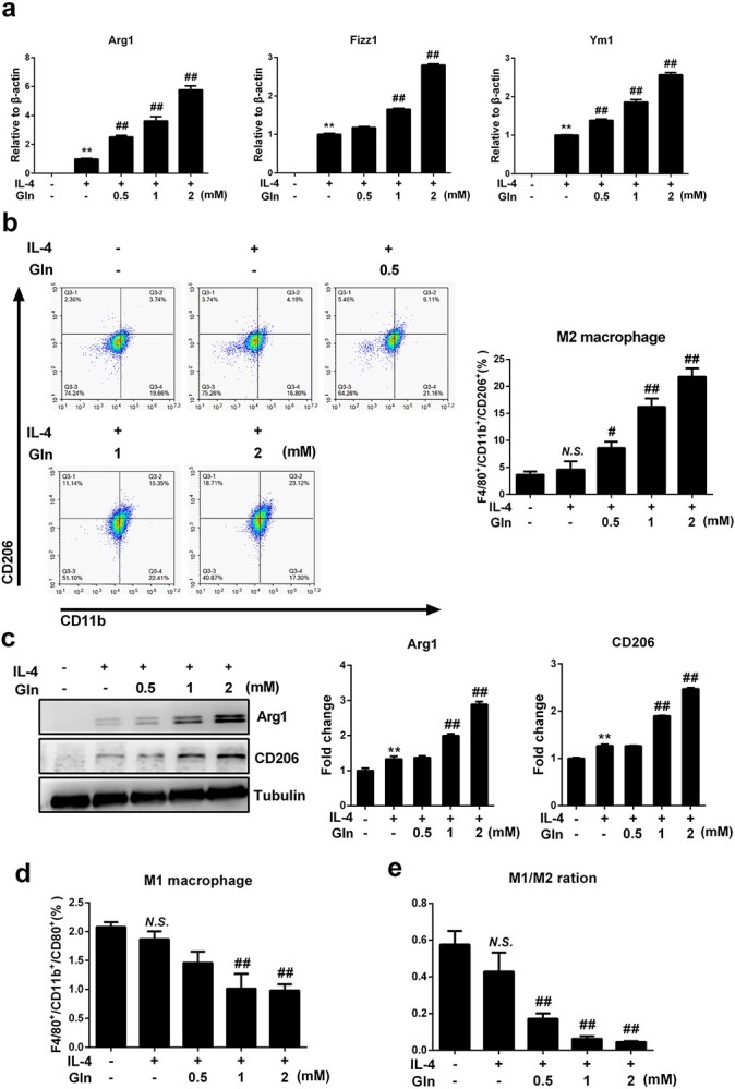
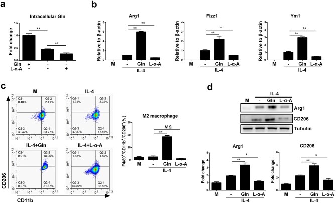
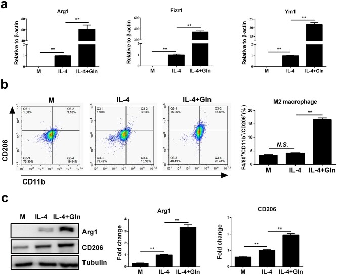
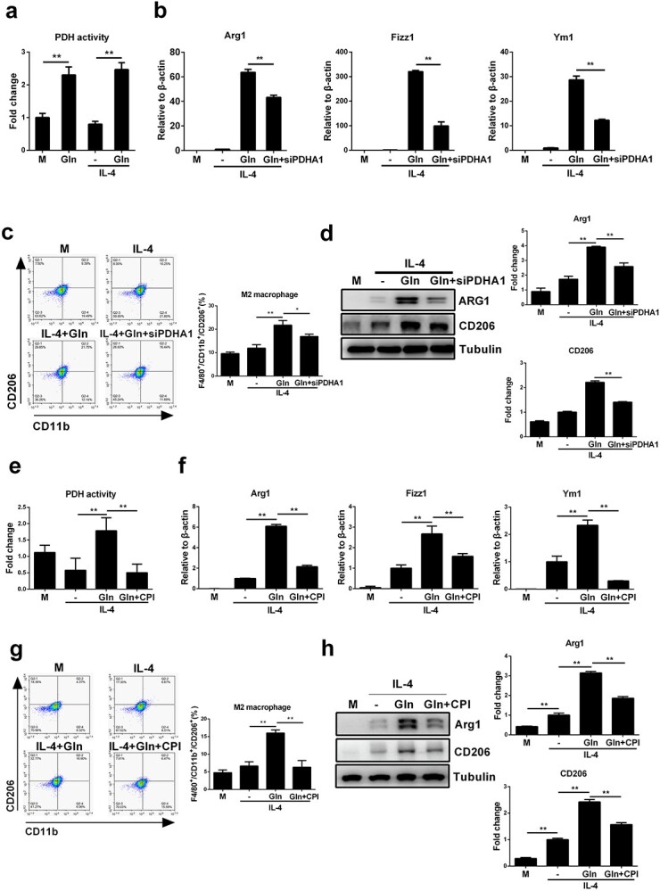
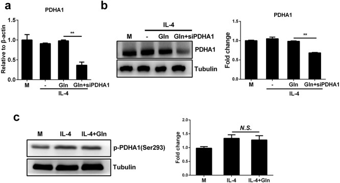
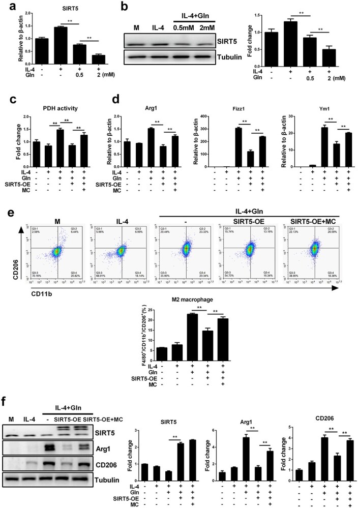
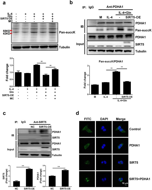
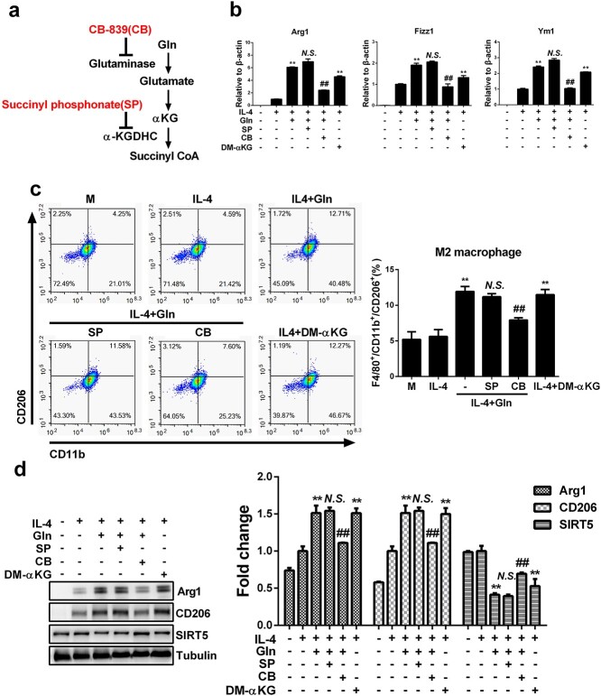
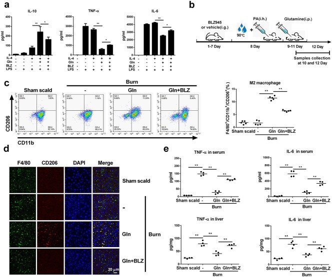
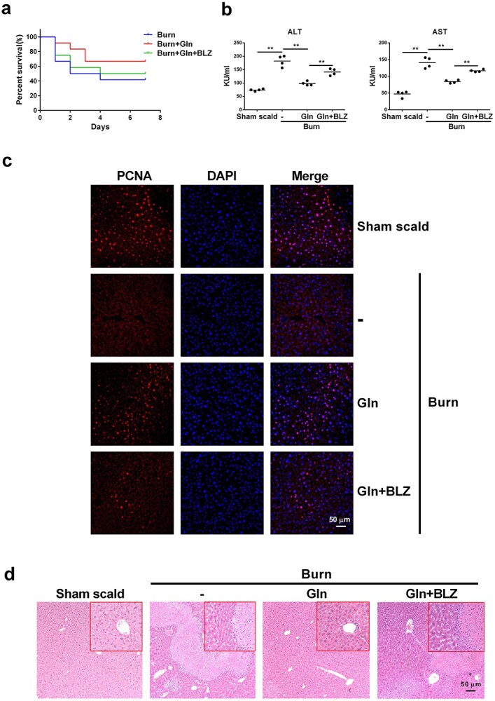
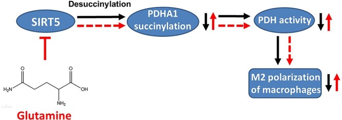
Results: We showed that consumption of glutamine supported M2 activation in IL-4-treated murine macrophages by upregulating the activity of PDH. Mechanistically, glutamine did not affect the expression or alter the phosphorylation status of PDHA1 but instead downregulated the expression of SIRT5 and repressed SIRT5-dependent desuccinylation on PDHA1, which in turn recovered PDH activity and supported M2 polarization. This effect was implemented by its secondary metabolite α-ketoglutarate (αKG) rather than glutamine itself. Finally, we demonstrated that glutamine promoted macrophage M2 polarization in a murine burn sepsis model, thereby repressing excessive inflammation and alleviating organ injury in model mice.

Conclusions: Glutamine mitigates murine burn sepsis by essentially supporting macrophage M2 polarization, with a mechanism involving the repression of the SIRT5-mediated desuccinylation of pyruvate dehydrogenase that replenishes OXPHOS and sustains M2 macrophages.

Keywords: Burn, Sepsis; Desuccinylation; Glutamine; Macrophages polarization; PDH; SIRT5.


Figures

None
Graphical abstract
Figure 1
Figure 1
Glutamine (Gln) supplement augments macrophage M2 polarization in IL-4 treated BMDMs. (a) BMDMs were treated with 50 ng/ml IL-4 alone or together with gradient concentrations of glutamine (Gln, 0.5–2 mM) for 24 h. mRNA expression of Arg1, Fizz1 and Ym1 was detected by RT-PCR (b). F4/80+/CD11b+/CD206+ cells were detected by flow cytometry (c). Protein expression of Arg1 and CD206 was detected by western blot. F4/80+/CD11b+/CD80+ cells were detected by flow cytometry (d). The ratio of M1 and M2 macrophages was detected by flow cytometry (e). **p < 0.01, N.S. no significance vs treatment without glutamine and IL-4. #p < 0.05, ##p < 0.01 vs IL-4 treatment alone. n = 3. BMDMs bone marrow derived macrophages, IL interleukin
Figure 2
Figure 2
Intracellular deprivation of glutamine (Gln) inhibits M2 polarization of macrophages. (a) BMDMs were supplemented with Gln (2 mM) or starved of Gln or further treated with L-α-Aminoadipic acid (L-α-A, 200 μM) for 24 h. Intracellular Gln was detected. (b) BMDMs were treated with IL-4 (50 ng/ml) alone or together with Gln (2 mM) or further with L-α-A (200 μM) for 24 h. mRNA expression of Arg1, Fizz1 and Ym1 was detected by RT-PCR. (c) F4/80+/CD11b+/CD206+ cells were detected by flow cytometry. (d) Protein expression of Arg1 and CD206 was detected by western blot. *p < 0.05, **p < 0.01. N.S. no significance. n = 3. BMDMs bone marrow derived macrophages, IL interleukin
Figure 3
Figure 3
Glutamine (Gln) supplementation augments macrophage M2 polarization in IL-4-treated J774A.1 cells. (a) J774A.1 cells were treated with 50 ng/ml IL-4 alone or together with 2 mM Gln for 24 h. mRNA expression of Arg1, Fizz1 and Ym1 was detected by RT-PCR. (b) F4/80+/CD11b+/CD206+ cells were detected by flow cytometry. (c) Protein expression of Arg1 and CD206 was detected by western blot. **p < 0.01. N.S. no significance. n = 3. IL interleukin
Figure 4
Figure 4
Glutamine (Gln) enhances macrophage M2 polarization by increasing the activity of PDH. (a) BMDMs were treated with 2mM Gln alone, 50ng/ml IL-4 alone or IL-4 together with Gln for 24 h. PDH activity was detected by absorbance detection. (b) J774A.1 cells were transfected with PDHA1 siRNA (siPDHA1) for 48 h and then treated with 50 ng/ml IL-4 alone or further with 2 mM Gln for 24 h. mRNA expression of Arg1, Fizz1 and Ym1 was detected by RT-PCR. (c) F4/80+/CD11b+/CD206+ cells were detected by flow cytometry. (d) Protein expression of Arg1 and CD206 was detected by western blot. (eh) BMDMs were treated with 50 ng/ml IL-4 alone or together with 2 mM Gln or further treated with 10 μM CPI-613 (CPI) for 24 h. PDH activity was detected by absorbance detection (e). mRNA expression of Arg1, Fizz1 and Ym1 was detected by RT-PCR (f). F4/80+/CD11b+/CD206+ cells were detected by flow cytometry (g). Protein expression of Arg1 and CD206 was detected by western blot (h). *p < 0.05, **p < 0.01. N.S., no significance. n = 3. BMDMs bone marrow derived macrophages, IL interleukin
Figure 5
Figure 5
Glutamine (Gln) promotes PDH activity without upregulating its expression or affecting its protein phosphorylation. (a,b) J774A.1 cells were transfected with PDHA1 siRNA (siPDHA1) for 48 h. Then cells were treated with 50 ng/ml IL-4 alone or together with 2 mM Gln for 24 h. mRNA (a) and protein (b) expression of PDHA1 were detected by RT-PCR and western blot, respectively. (c) BMDMs were treated with 50 ng/ml IL-4 alone or together with 2 mM Gln for 24 h. The phosphorylation of PDHA1 (p-PDHA1) was detected by western blot. **p < 0.01. N.S. no significance. n = 3. BMDMs bone marrow derived macrophages, IL interleukin
Figure 6
Figure 6
Glutamine (Gln) promotes PDH activity and enhances macrophages M2 polarization by inhibiting SIRT5. (a,b) Control J774A.1 cells and SIRT5-overexpressing (SIRT5-OE) J774A.1 cells were treated with 50 ng/ml IL-4 alone or together with Gln (0.5 and 2 mM) for 24 h. mRNA (a) and protein (b) levels of SIRT5 were detected by RT-PCR and western blot, respectively. (c) Control J774A.1 cells and SIRT5-OE J774A.1 cells were treated with 50 ng/ml IL-4 alone or further with 2 mM Gln or 50 μM MC3482 (MC) for 24 h. PDH activity was detected by absorbance detection. (df) Control J774A.1 cells and SIRT5-OE J774A.1 cells were treated with 50 ng/ml IL-4 alone or together with 2 mM Gln or further treated with 50 μM MC3482 (MC) for 24 h. mRNA expression of Arg1, Fizz1 and Ym1 was detected by RT-PCR (d). F4/80+/CD11b+/CD206+ cells were detected by flow cytometry (e). Protein expression of SIRT5, Arg1 and CD206 was detected by western blot (f). **p < 0.01. n = 3. IL interleukin
Figure 7
Figure 7
Glutamine (Gln) regulated PDHA1 succinylation modification by inhibiting the desuccinylation modification activity of SIRT5. (a) Control J774A.1 cells and SIRT5-OE J774A.1 cells were treated with 50 ng/ml IL-4 or IL-4 with 2 mM Gln or further treated with 50 μM MC3482 (MC) for 24 h. Pan-succinyllysine (Pan-succK) was detected by western blot. (b) Control J774A.1 cells and SIRT5-OE J774A.1 cells were treated with 50 ng/ml IL-4 or IL-4 with 2 mM Gln. PDHA1 protein was precipitated by immunoprecipitation using PDHA1 antibody and the succinylation modification of PDHA1 was detected by western blot using a Pan-succK antibody. (c) The amount of PDHA1 conjugated with SIRT5 was detected by co-immunoprecipitation assay. (d) pBIFC-VN173-PDHA1 (PDHA1) and pBIFC-VC155-SIRT5 (SIRT5) plasmids were transfected separately or together into 293 T cells for 48 h. Fluorescence images were detected by Ziss LSM880 laser scanning confocal microscope. Scale bar, 20 µm. **p < 0.01. n = 3. IL interleukin
Figure 8
Figure 8
Glutamine (Gln) increases macrophage M2 polarization by its metabolite α-ketoglutarate (αKG). (a) A metabolic flowchart of Gln and the targets of inhibitors. (bd) BMDMs were treated with 50 ng/ml IL-4, IL-4 with 2 mM dimethyl α-ketoglutarate (DM-αKG) or IL-4 with 2 mM Gln and further treated with 10 μM CB-839 (CB) or 50 μM succinyl phosphonate (SP) for 24 h. mRNA expression of Arg1, Fizz1 and Ym1 was detected by RT-PCR (b). F4/80+/CD11b+/CD206+ cells were detected by flow cytometry (c). Protein expression of Arg1, CD206 and SIRT5 was detected by western blot (d). **P < 0.01 vs IL-4, ##p < 0.01, N.S., no significance vs IL-4 + Gln. n = 3. BMDMs bone marrow derived macrophages, IL interleukin
Figure 9
Figure 9
Glutamine (Gln) promoted M2 polarization of macrophages and reduced inflammation in burn sepsis mice. (a) Murine BMDMs were treated with 50 ng/ml IL-4, or IL-4 with 2 mM Gln or further treated with 10 μM BLZ-945 (BLZ) for 24 h. Then cells were stimulated with 20 ng/ml LPS for 24 h. Supernatant IL-10, TNF-α and IL-6 were detected by ELISA (n = 3). (be) Wild-type BALB/c mice were pretreated with BLZ945 (BLZ) for 7 days, then intraperitoneally injected with normal saline (Burn) or 0.75 g/kg Gln every 24 h after the burn sepsis model was created. Serum, peritoneal fluid and liver tissue were collected 1 day after Gln injection (b). F4/80+/CD11b+/CD206+ cells in peritoneal fluid were detected by flow cytometry (c) (n = 5). Immunofluorescence staining of liver tissue for F4/80 and CD206 was detected by ZISS LSM880 laser scanning confocal microscope (d). TNF-α and IL-6 in serum and liver tissue homogenate were detected by ELISA assay (e) (n = 4). **p < 0.01. Scale bar, 20 μm. i.g. intragastric administration, i.h. hypodermic injection, i.p. intraperitoneal injection. BMDMs bone marrow derived macrophages, IL interleukin, TNF-α tumor necrosis factor-α, LPS lipopolysaccharide
Figure 10
Figure 10
Glutamine (Gln) alleviates burn sepsis and organ damage. (ad) Wild-type BALB/c mice were pretreated consecutively with BLZ945 (BLZ) for 7 days, then intraperitoneally injected with normal saline (Burn) or 0.75 g/kg Gln 24 h after the burn sepsis model was created. The survival of the mice was observed for 7 days (a) (n = 10). Serum was collected 1 day after Gln injection and the levels of ALT and AST were detected (b) (n = 4). Liver tissues were collected 3 days after Gln injection. The immunofluorescence of proliferating cell nuclear antigen (PCNA) in liver tissue was detected (c). Histopathological examination of liver was performed by hematoxylin and oesin (HE) staining (d). **p < 0.01. N.S. no significance. Scale bar, 50 μm. ALT alanine transaminase, AST aspartate aminotransferase
Figure 11
Figure 11
Schematic model depicting a hypothetical mechanism of glutamine promoting M2 polarization in macrophages

Similar articles

Cited by

References

    1. Zhang P, Zou B, Liou YC, Huang C. The pathogenesis and diagnosis of sepsis post burn injury. Burns Trauma. 2021;9:tkaa047. 10.1093/burnst/tkaa047. - DOI - PMC - PubMed
    1. Kamolz LP. Burns: learning from the past in order to be fit for the future. Crit Care. 2010;14:106. 10.1186/cc8192. - DOI - PMC - PubMed
    1. Stanojcic M, Vinaik R, Abdullahi A, Chen P, Jeschke MG. NLRP3 knockout enhances immune infiltration and inflammatory responses and improves survival in a burn sepsis model. Immunology. 2022;165:195–205. - PMC - PubMed
    1. Locati M, Curtale G, Mantovani A. Diversity, mechanisms, and significance of macrophage plasticity. Annu Rev Pathol. 2020;15:123–47. - PMC - PubMed
    1. Luck ME, Herrnreiter CJ, Choudhry MA. Gut microbial changes and their contribution to post-burn pathology. Shock. 2021;56:329–44. - PMC - PubMed