. 2022 Dec 19:10:989932.
doi: 10.3389/fbioe.2022.989932. eCollection 2022.

Novel assay to measure chromosome instability identifies Punica granatum extract that elevates CIN and has a potential for tumor- suppressing therapies

Affiliations

Novel assay to measure chromosome instability identifies Punica granatum extract that elevates CIN and has a potential for tumor- suppressing therapies

Nikolay V Goncharov et al. Front Bioeng Biotechnol. .

Abstract

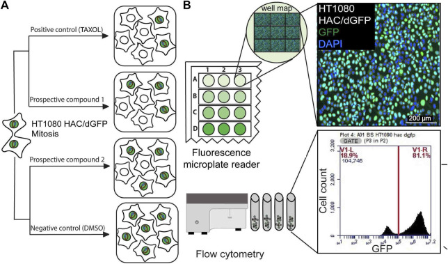
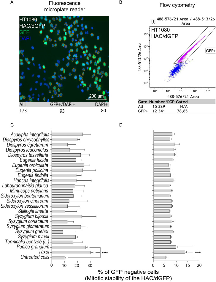
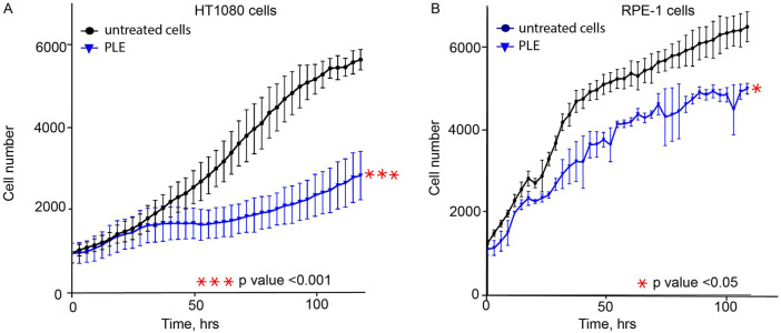
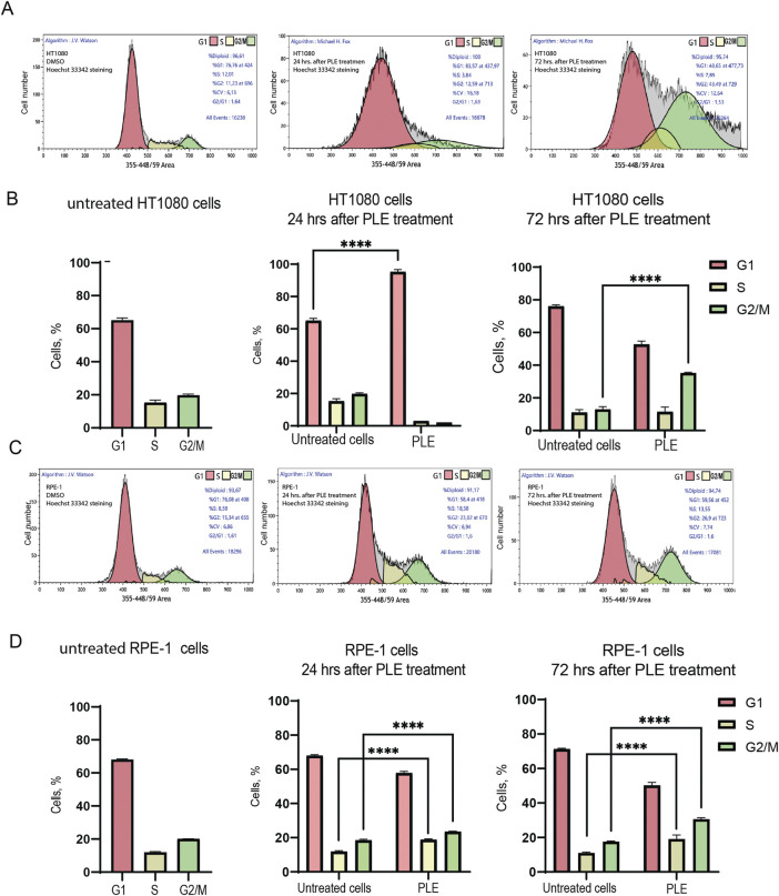
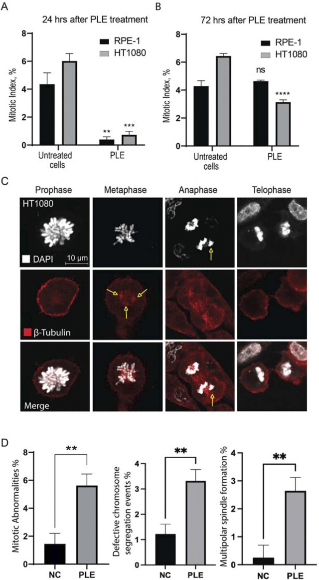
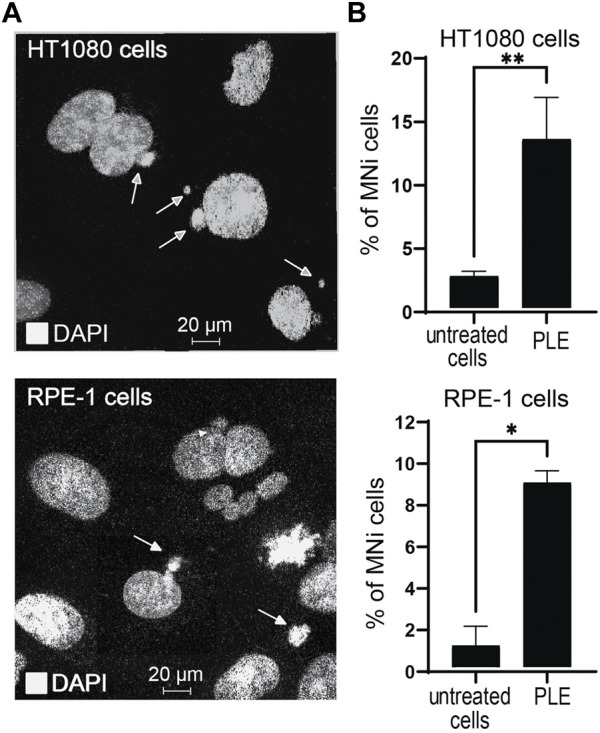
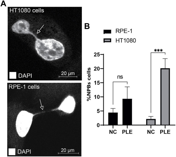
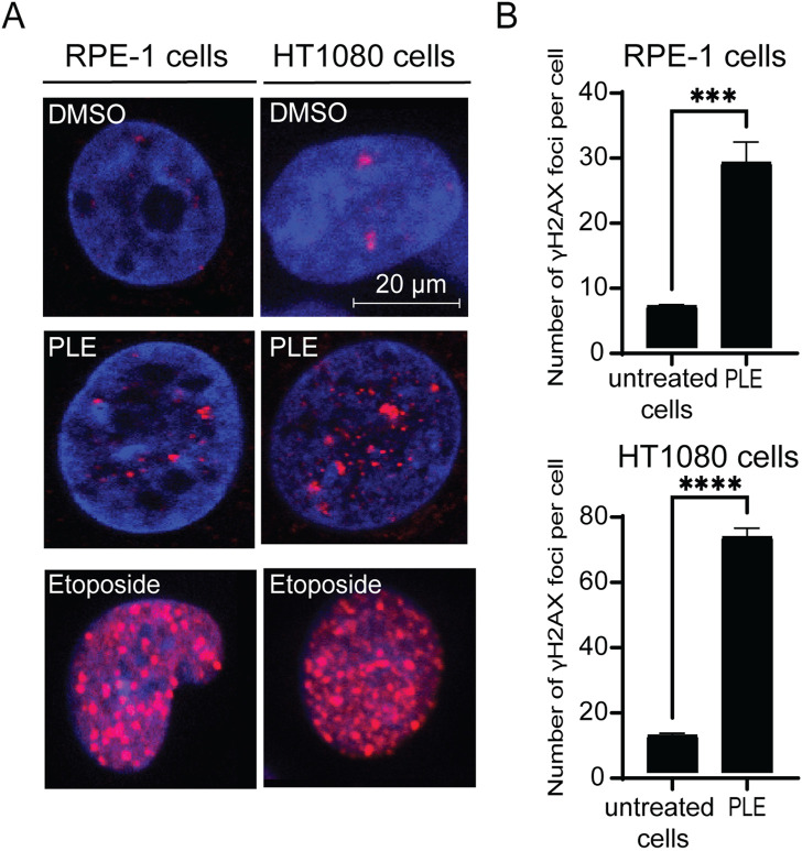
Human artificial chromosomes (HACs) have provided a useful tool to study kinetochore structure and function, gene delivery, and gene expression. The HAC propagates and segregates properly in the cells. Recently, we have developed an experimental high-throughput imaging (HTI) HAC-based assay that allows the identification of genes whose depletion leads to chromosome instability (CIN). The HAC carries a GFP transgene that facilitates quantitative measurement of CIN. The loss of HAC/GFP may be measured by flow cytometry or fluorescence scanning microscope. Therefore, CIN rate can be measured by counting the proportion of fluorescent cells. Here, the HAC/GFP-based assay has been adapted to screen anticancer compounds for possible induction or elevation of CIN. We analyzed 24 cytotoxic plant extracts. Punica granatum leaf extract (PLE) indeed sharply increases CIN rate in HT1080 fibrosarcoma cells. PLE treatment leads to cell cycle arrest, reduction of mitotic index, and the increased numbers of micronuclei (MNi) and nucleoplasmic bridges (NPBs). PLE-mediated increased CIN correlates with the induction of double-stranded breaks (DSBs). We infer that the PLE extract contains a component(s) that elevate CIN, making it a candidate for further study as a potential cancer treatment. The data also provide a proof of principle for the utility of the HAC/GFP-based system in screening for natural products and other compounds that elevate CIN in cancer cells.

Keywords: CIN; HAC; Punica granatum leave extract; chromosome instability; double-stranded breaks; human artificial chromosome; micronuclei; natural products.


Conflict of interest statement

The authors declare that the research was conducted in the absence of any commercial or financial relationships that could be construed as a potential conflict of interest.

Figures

FIGURE 1
FIGURE 1
(A) A scheme of the HAC/dGFP assay. The assay is based on the use of the HAC/dGFP (the alphoidtetO-HAC containing the dGFP transgene). Cells with the HAC display green fluorescence, while cells that lack it do not. It is expected that untreated cells (“a negative control”) should show uniform green fluorescence. (B) A cell population, that has lost HAC upon treatment with the CIN-inducing drug (“a prospective compound”), loses GFP fluorescence. For the positive control, we used Taxol, a CIN-inducing microtubule destabilizing agent. The actual number and percentage of cells with the HAC/dGFP are measured by a fluorescence microplate reader and flow cytometry.
FIGURE 2
FIGURE 2
Mitotic stability of the HAC/dGFP in human cancer HT1080 cells treated with the natural extracts. Mitotic stability was measured by a fluorescent microplate reader (A,C) and flow cytometry (B,D). Among 24 extracts analyzed, the strongest effect on the HAC/dGFP stability was demonstrated by the cells treated with the Punica granatum leaves extract (PLE). Asterisks indicate statistical significance above negative control (T-test, p < 0.01).
FIGURE 3
FIGURE 3
The proliferation rate of HT1080 (A) and RPE-1 (B) cells treated with PLE. Cells were treated for 24 h, then media was replaced, and cell proliferation was recorded by live imaging for 120 h. PLE—P. granatum leaves. Each cell line was compared to a negative control: HT1080 PLE-treated cells compared to HT1080 untreated cells; RPE-1 PLE-treated cells compared to RPE-1 untreated cells. The statistical analysis was performed using an unpaired Student's t-test. Asterisks indicate statistical significance (*p < 0.05, ***p < 0.001, relative to the negative control – untreated cells).
FIGURE 4
FIGURE 4
Analysis of the cell cycle after PLE treatment. (A) Flow cytometry analysis of HT1080 untreated cells 24 and 72 h after PLE treatment. (B) The percentage of cells in G1, S and G2/M phases in HT1080 cell population after 24 and 72 h. (C) Flow cytometry analysis of RPE-1 untreated cells after 24 and 72 h. (D) The percentage of cells in G1, S and G2/M phases in RPE-1 cell population after 24 and 72 h. The statistical analysis was performed using 2way ANNOVA, asterisks indicate statistical significance (****p < 0,0001, relative to the negative control—untreated cells).
FIGURE 5
FIGURE 5
Mitotic index of HT1080 and RPE-1 cells treated with PLE. (A) The percentage of mitosis in HT1080 and RPE-1cells 24 h after PLE treatment. (B) The percentage of mitosis 72 h after PLE treatment. (C) Analysis of localization of tubulin beta at the different stages of mitosis after PLE treatment in HT1080 cells. Staining by antibodies against tubulin beta is marked in red and DAPI in white. Yellow arrows point to the observed mitotic abnormalities. (D) Proportion of observed mitotic abnormalities in HT1080 cells. Representative pictures of localization of tubulin beta at the different stages of mitosis in untreated HT1080 cells are shown in Supplementary Figure S4. Between 100 and 150 mitotic events were analyzed. Statistical analysis was performed using 2way ANNOVA, asterisks indicate statistical significance (**p < 0.01, ***p < 0.001, ****p < 0.0001 relative to the negative control—untreated cells).
FIGURE 6
FIGURE 6
Micronuclei (MNi) formation 24 h after PLE treatment of HT1080 and RPE-1cells. (A) Examples of (MNi) formation in HT1080 and RPE-1 cells. White arrows point to the MNi. (B) The percentage of MNi after PLE treatment compared to untreated cells. Error bars correspond to a standard deviation (SD) of four replicates. Statistical analysis was performed using one-way ANNOVA, asterisks indicate statistical significance (*p < 0.05, **p < 0.01, relative to the negative control—untreated cells).
FIGURE 7
FIGURE 7
Accumulation of NPBs in HT1080 and RPE-1 cells treated with PLE. (A) Representative pictures of NPBs in HT1080 and RPE-1 cells after PLE treatment. (B) The percentage of NPBs in HT1080 and RPE-1 cells. Error bars correspond to a standard deviation (SD) of four replicates. Statistical analysis was performed using 2way ANNOVA, asterisks indicate statistical significance (*p < 0.005, ***p < 0.0005, relative to the negative control—untreated cells).
FIGURE 8
FIGURE 8
Accumulation of γH2AX foci in HT1080 and RPE-1 cells treated with PLE. (A) γH2AX immunostaining of HT1080 cells before and after treatment with PLE. (B) The percentage of γH2AX foci in HT1080 and RPE-1 cells. As a positive control, we used Etoposide – a drug commonly used for cancer therapy. Etoposide is an inhibitor of topoisomerase II that forms DSBs (Tassanee Lerksuthirat et all., 2020). Error bars correspond to a standard deviation (SD) of four replicates. Statistical analysis was performed using 2way ANNOVA, asterisks indicate statistical significance (***p < 0.001, ****p < 0.0001, relative to the negative control—untreated cells).

References

    1. Ahmed M. B., A Alghamdi A. A., Islam S. U., Ahsan H., Lee Y. S. (2022). The complex roles of DNA repair pathways, inhibitors, hyperthermia, and contact inhibition in cell cycle halts. Mini Rev. Med. Chem. [Epub ahead of print] 22. 10.2174/1389557522666220826141837 - DOI - PubMed
    1. Alberto Coronado-Reyes J., CortÉS-Penagos C., González-Hernández J. (2021). Chemical composition and great applications to the fruit of the pomegranate (Punica granatum): a review. Food Sci. Technol. 42. 10.1590/fst.29420 - DOI
    1. Bakker B., van den Bos H., Lansdorp P. M., Foijer F. (2015). How to count chromosomes in a cell: An overview of current and novel technologies. Bioessays 37 (5), 570–577. 10.1002/bies.201400218 - DOI - PubMed
    1. Cai Y. Z., Mei S., Jie X., Luo Q., Corke H. (2006). Structure-radical scavenging activity relationships of phenolic compounds from traditional Chinese medicinal plants. Life Sci. 78 (25), 2872–2888. 10.1016/j.lfs.2005.11.004 - DOI - PubMed
    1. Costa-Lotufo L. V., Khan M. T., Ather A., Wilke D. V., Jimenez P. C., Pessoa C., et al. (2005). Studies of the anticancer potential of plants used in Bangladeshi folk medicine. J. Ethnopharmacol. 99 (1), 21–30. 10.1016/j.jep.2005.01.041 - DOI - PubMed

LinkOut - more resources