. 2022 Dec 22;4(6):e220155.
doi: 10.1148/ryct.220155. eCollection 2022 Dec.

Limited Aortic Intimal Tears: CT Imaging Features and Clinical Characteristics

Affiliations

Limited Aortic Intimal Tears: CT Imaging Features and Clinical Characteristics

Mohammad H Madani et al. Radiol Cardiothorac Imaging. .

Abstract

Limited aortic intimal tear is an uncommon lesion of the dissection spectrum. The lesion has several imaging features that are not well known, including asymmetric aortic contour abnormalities, filling defects, and various morphologic patterns, such as linear, L-shaped, T-shaped, and stellate configurations. Hemorrhage of the aortic wall may also be present in patients with this rare entity. This imaging essay reviews the CT imaging findings and clinical characteristics of patients with limited intimal tears. Keywords: Aorta, CT © RSNA, 2022.

Keywords: Aorta; CT.


Conflict of interest statement

Disclosures of conflicts of interest: M.H.M No relevant relationships. V.L.T. Shareholder of Segmed. R.L.H. No relevant relationships. M.J.W. Postdoctoral fellowship grant from the American Heart Association. H.M. No relevant relationships. A.S.C. No relevant relationships. G.J.B. No relevant relationships. D.F. Deputy editor for Radiology: Cardiothoracic Imaging.

Figures
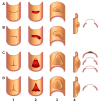
Drawings of limited aortic intimal tear configurations. (A) Linear, (B)
L-shaped, (C) T-shaped, and (D) stellate tear configurations within endoluminal
(1–2) and external surface (3–4) views of the aorta. Linear tear
represented a tear in a single plane or orientation. L-shaped tear reflected a
linear tear with an additional tear plane oriented at the end and perpendicular
to the linear tear. T-shaped tear had an additional tear plane oriented at the
mid segment and perpendicular to a linear tear plane. Stellate tear represented
a star-shaped configuration.
Figure 1:
Drawings of limited aortic intimal tear configurations. (A) Linear, (B) L-shaped, (C) T-shaped, and (D) stellate tear configurations within endoluminal (1–2) and external surface (3–4) views of the aorta. Linear tear represented a tear in a single plane or orientation. L-shaped tear reflected a linear tear with an additional tear plane oriented at the end and perpendicular to the linear tear. T-shaped tear had an additional tear plane oriented at the mid segment and perpendicular to a linear tear plane. Stellate tear represented a star-shaped configuration.
Linear-shaped limited intimal tear (LIT). (A) Blood-pool inversion
volume-rendered endoluminal CT image shows ovoid, linear type A LIT in the
aortic root (arrowheads). Note proximity to the left (L) and right (R) coronary
arteries. (B) Volume-rendered CT image of external surface of the aortic root
demonstrates linear defect (arrowheads) spanning the distance between the left
(L) and right (R) coronary arteries. (C) Axial contrast-enhanced CT angiographic
image of the ascending thoracic aorta (Ao) shows focal linear filling defects
(arrowheads) representing the undermined edges of the tear.
Figure 2:
Linear-shaped limited intimal tear (LIT). (A) Blood-pool inversion volume-rendered endoluminal CT image shows ovoid, linear type A LIT in the aortic root (arrowheads). Note proximity to the left (L) and right (R) coronary arteries. (B) Volume-rendered CT image of external surface of the aortic root demonstrates linear defect (arrowheads) spanning the distance between the left (L) and right (R) coronary arteries. (C) Axial contrast-enhanced CT angiographic image of the ascending thoracic aorta (Ao) shows focal linear filling defects (arrowheads) representing the undermined edges of the tear.
L-shaped limited intimal tear (LIT). (A) Blood-pool inversion
volume-rendered endoluminal CT image shows type A LIT with L-shaped
configuration in the ascending thoracic aorta (arrowheads). (B) Volume-rendered
CT image of external surface of ascending aorta shows the tear (arrowheads). (C)
Axial contrast-enhanced CT angiographic image demonstrates undermined edges of
the tear (arrowheads), with focal outpouching of the remaining aortic wall
(**).
Figure 3:
L-shaped limited intimal tear (LIT). (A) Blood-pool inversion volume-rendered endoluminal CT image shows type A LIT with L-shaped configuration in the ascending thoracic aorta (arrowheads). (B) Volume-rendered CT image of external surface of ascending aorta shows the tear (arrowheads). (C) Axial contrast-enhanced CT angiographic image demonstrates undermined edges of the tear (arrowheads), with focal outpouching of the remaining aortic wall (**).
T-shaped limited intimal tear (LIT). (A) Blood-pool inversion
volume-rendered endoluminal CT image shows type A LIT with T-shaped tear vector,
resulting in a triangular tear pattern (arrowheads). (B) Volume-rendered CT
image of external surface shows the tear (arrowheads). (C) Volume-rendered CT
image shows focal outpouching between the undermined edges of the LIT
(arrowheads). (D) Axial contrast-enhanced CT angiographic image demonstrates
elongated undermined edges (arrowheads). A portion of an associated intramural
hematoma is also demonstrated (**).
Figure 4:
T-shaped limited intimal tear (LIT). (A) Blood-pool inversion volume-rendered endoluminal CT image shows type A LIT with T-shaped tear vector, resulting in a triangular tear pattern (arrowheads). (B) Volume-rendered CT image of external surface shows the tear (arrowheads). (C) Volume-rendered CT image shows focal outpouching between the undermined edges of the LIT (arrowheads). (D) Axial contrast-enhanced CT angiographic image demonstrates elongated undermined edges (arrowheads). A portion of an associated intramural hematoma is also demonstrated (**).
Stellate-shaped limited intimal tear (LIT). (A) Blood-pool inversion
volume-rendered endoluminal CT image shows type A LIT with stellate tear vector,
resulting in large ovoid defect (arrowheads). (B) Volume-rendered CT image of
external surface shows large ovoid bulge (arrowheads) representing the remaining
media and adventitia within the LIT. (C) Axial contrast-enhanced CT angiographic
image demonstrates undermined edges (arrows) at the LIT in the proximal
transverse aorta (Ao). Large bulge anteriorly between the edges represents the
residual aortic wall (arrowheads).
Figure 5:
Stellate-shaped limited intimal tear (LIT). (A) Blood-pool inversion volume-rendered endoluminal CT image shows type A LIT with stellate tear vector, resulting in large ovoid defect (arrowheads). (B) Volume-rendered CT image of external surface shows large ovoid bulge (arrowheads) representing the remaining media and adventitia within the LIT. (C) Axial contrast-enhanced CT angiographic image demonstrates undermined edges (arrows) at the LIT in the proximal transverse aorta (Ao). Large bulge anteriorly between the edges represents the residual aortic wall (arrowheads).
Limited intimal tear (LIT) of descending thoracic aorta with intramural
hematoma. (A) Blood-pool inversion volume-rendered endoluminal CT image shows
type B LIT (arrowheads) of the proximal descending thoracic aorta. (B)
Volume-rendered CT image of external surface shows the tear (arrowheads). (C)
Axial contrast-enhanced CT angiographic image of type B LIT (arrowheads) and
intramural hematoma (**).
Figure 6:
Limited intimal tear (LIT) of descending thoracic aorta with intramural hematoma. (A) Blood-pool inversion volume-rendered endoluminal CT image shows type B LIT (arrowheads) of the proximal descending thoracic aorta. (B) Volume-rendered CT image of external surface shows the tear (arrowheads). (C) Axial contrast-enhanced CT angiographic image of type B LIT (arrowheads) and intramural hematoma (**).
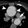
Limited intimal tears (LITs) and intramural hematoma (IMH). Axial
contrast-enhanced image from CT angiography demonstrates LIT with focal bulge in
the ascending thoracic aorta (arrowheads), as well as LIT (arrow) and IMH
(**) in the descending thoracic aorta. IMH was hyperattenuated on
noncontrast imaging (not shown).
Figure 7:
Limited intimal tears (LITs) and intramural hematoma (IMH). Axial contrast-enhanced image from CT angiography demonstrates LIT with focal bulge in the ascending thoracic aorta (arrowheads), as well as LIT (arrow) and IMH (**) in the descending thoracic aorta. IMH was hyperattenuated on noncontrast imaging (not shown).
Ancillary findings of hemopericardium (mean attenuation, 46 HU) and
bilateral hemothorax (mean attenuation, 28 HU) (arrowheads) on axial noncontrast
CT image in a patient with limited intimal tear of the ascending thoracic
aorta.
Figure 8:
Ancillary findings of hemopericardium (mean attenuation, 46 HU) and bilateral hemothorax (mean attenuation, 28 HU) (arrowheads) on axial noncontrast CT image in a patient with limited intimal tear of the ascending thoracic aorta.
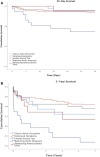
Thirty-day and 5-year survival rates in patients with acute aortic
syndromes.
Figure 9:
Thirty-day and 5-year survival rates in patients with acute aortic syndromes.

References

    1. Murray CA , Edwards JE . Spontaneous laceration of ascending aorta . Circulation 1973. ; 47 ( 4 ): 848 – 858 . - PubMed
    1. Widder DJ , Novelline RA , Derkac WM . Spontaneous nontraumatic rupture of the thoracic aorta . J Thorac Cardiovasc Surg 1983. ; 86 ( 4 ): 626 – 628 . - PubMed
    1. Padró JM , Caralps JM , García J , Arís A . Spontaneous rupture of the ascending aorta . J Cardiovasc Surg (Torino) 1988. ; 29 ( 1 ): 109 – 110 . - PubMed
    1. Aoyagi S , Akashi H , Fujino T , et al. . Spontaneous rupture of the ascending aorta . Eur J Cardiothorac Surg 1991. ; 5 ( 12 ): 660 – 662 . - PubMed
    1. Handa N , Takamoto S , Hatanaka M , et al. . Spontaneous non-traumatic rupture of the thoracic aorta . Thorac Cardiovasc Surg 1994. ; 42 ( 6 ): 355 – 357 . - PubMed