. 2023 Jan 1;79(Pt 1):50-65.
doi: 10.1107/S2059798322011871. Epub 2023 Jan 1.

Molecular-dynamics simulation methods for macromolecular crystallography

Affiliations

Molecular-dynamics simulation methods for macromolecular crystallography

David C Wych et al. Acta Crystallogr D Struct Biol. .

Abstract

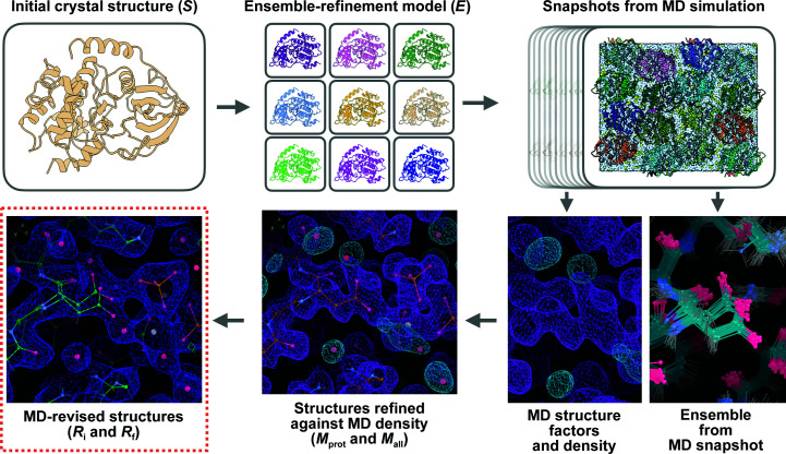
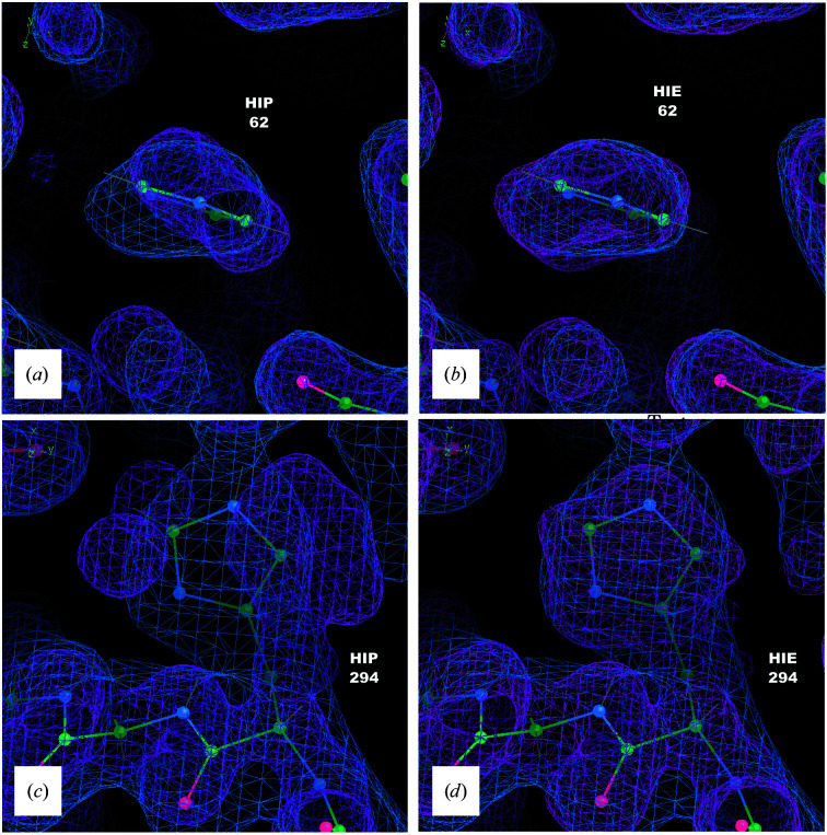
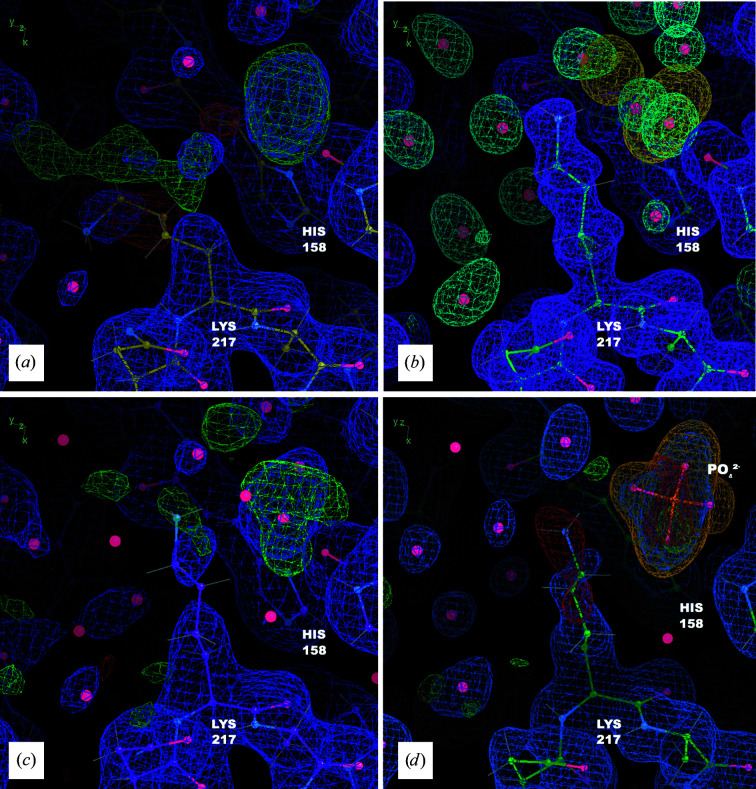
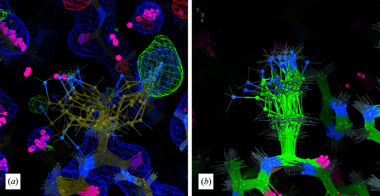
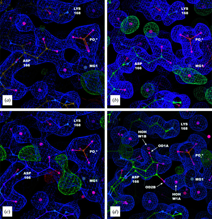
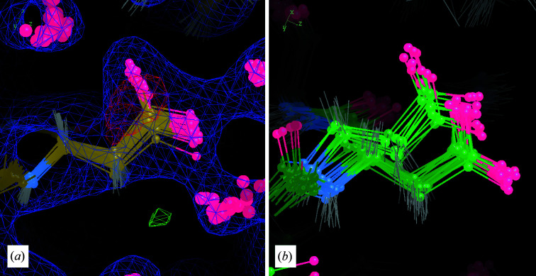
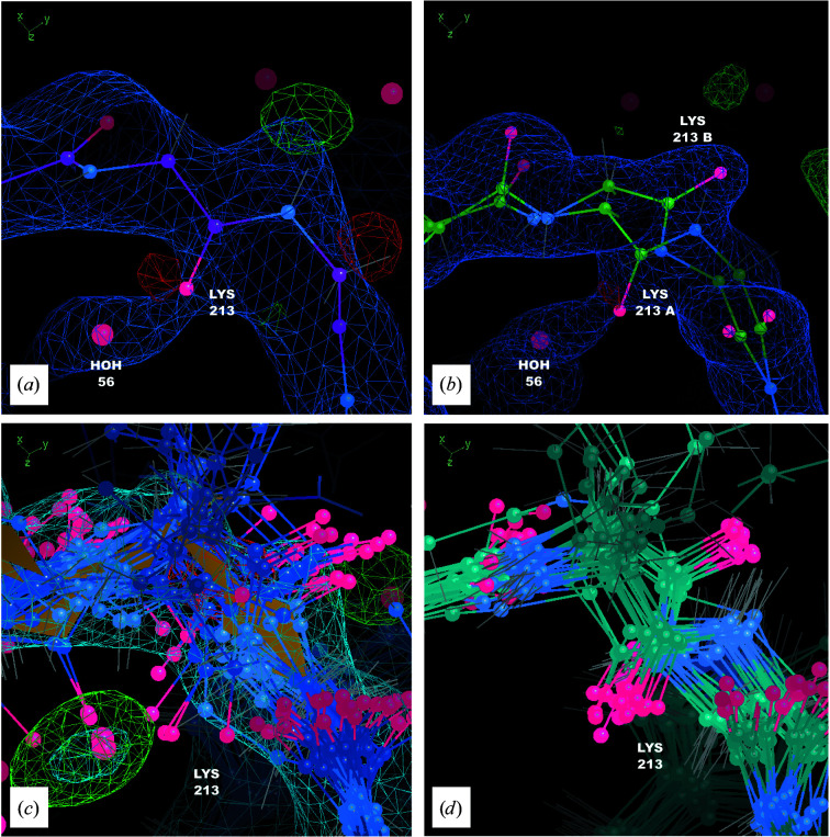
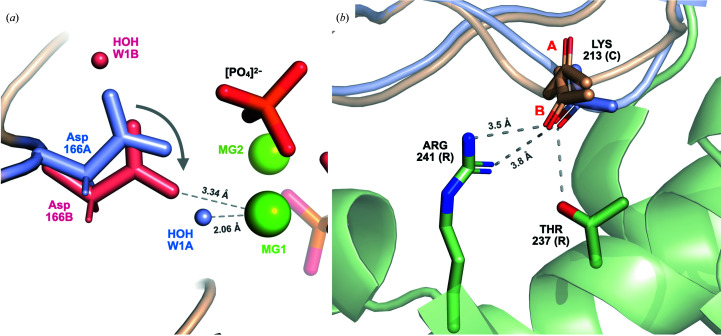
It is investigated whether molecular-dynamics (MD) simulations can be used to enhance macromolecular crystallography (MX) studies. Historically, protein crystal structures have been described using a single set of atomic coordinates. Because conformational variation is important for protein function, researchers now often build models that contain multiple structures. Methods for building such models can fail, however, in regions where the crystallographic density is difficult to interpret, for example at the protein-solvent interface. To address this limitation, a set of MD-MX methods that combine MD simulations of protein crystals with conventional modeling and refinement tools have been developed. In an application to a cyclic adenosine monophosphate-dependent protein kinase at room temperature, the procedure improved the interpretation of ambiguous density, yielding an alternative water model and a revised protein model including multiple conformations. The revised model provides mechanistic insights into the catalytic and regulatory interactions of the enzyme. The same methods may be used in other MX studies to seek mechanistic insights.

Keywords: conformational ensembles; molecular-dynamics simulations; protein kinases; water structure.


Figures

Figure 1
Figure 1
MD–MX procedure to revise macromolecular crystal structures. In this procedure, an initial crystal structure S (top left) is used to prepare an MD simulation model, using ensemble refinement to generate a set of diverse conformations (the ensemble-refinement model, E; top center). Snapshots from the MD simulation (top right) are used to generate simulated data and MD ensemble visualizations (bottom right; protein density in purple and water density in cyan). The initial structure is refined against the simulated data, leading to a revised protein structure, M prot, and a new water network, M all (bottom center; densities on bottom right). The revised structure is refined against the experimental data to produce the initial MD-revised structure R i, and manual improvements are made guided by the MD density and snapshots, leading to a final MD-revised structure R f (bottom left; 2F oF c density in blue at 1σ with traces of positive 3σ F oF c density visible in green).
Figure 2
Figure 2
The His62 and His294 MD densities agree with the crystallographic density when the residues are protonated at the ɛ atom, but disagree when they are doubly protonated. Coordinates of S are shown as sticks with 2F o − F c density in blue (1σ isosurface) and the total density from a 90–100 ns segment of a 200 kJ mol−1 nm−2 MD simulation in pink (1σ isosurface). (a) His62: MD density from a doubly protonated (HIP) simulation. (b) His62: MD density from simulating with histidine singly protonated (on the ɛ N atom; HIE). (c) His294: MD density from a doubly protonated (HIP) simulation. (d) His294: MD density from simulating with histidine singly protonated (on the ɛ N atom; HIE).
Figure 3
Figure 3
The MD–MX procedure guides remodeling of Lys217 and reveals a bound inorganic phosphate. (a) Coordinates, 2F oF c density (blue, 1σ isosurface) and F oF c density (positive in green and negative in red, 3σ isosurface) from S. (b) Coordinates from M all, with MD protein (purple, 1σ isosurface), solvent (blue, 3σ isosurface) and chloride density (yellow, 10σ isosurface), from the 90–100 ns segment of the simulation; the MD simulation suggests a different conformation for the side chain and a number of ordered waters; it also includes a spot of chloride density in the same position as the positive difference density in (a). (c) Coordinates, 2F oF c density (blue, 1σ isosurface) and F oF c density (positive in green and negative in red, 3σ isosurface) from R i; the shape of the difference density is suggestive of a coordinated free phosphate molecule. (d) Coordinates, 2F oF c density (blue, 1σ isosurface) and F oF c density (positive in green and negative in red, 3σ isosurface) from model R f refined against the high-resolution data (PDB entry 7v0g): the revised side-chain conformation, water network and phosphate are plausible and improve the difference density in the region. His158 is also shown as a reference point. Polder OMIT map density for the phosphate is shown at a level of 5σ (orange) confirming that the addition of this molecule is reasonable.
Figure 4
Figure 4
An MD snapshot guides remodeling of Lys217. (a) Coordinates, 2F oF c density (blue, 1σ isosurface) and F o − F c density (positive in green, negative in red, 3σ isosurface) from the ensemble-refinement model E: the Lys217 amino group is diverse and includes extensions into off-backbone density and positive difference density where the phosphate was placed in the R f structure (Fig. 3 ▸). (b) Final-frame MD snapshot: the side-chain conformations are more tightly clustered, extending straight from the backbone.
Figure 5
Figure 5
The MD–MX procedure yields a multi-conformer model of Asp166. (a) Coordinates, 2F oF c density (blue, 1σ isosurface) and F oF c density (positive in green and negative in red, 3σ isosurface) from model S. (b) Coordinates from the M all model, with MD protein and cofactor density (purple, 1σ isosurface) and solvent density (blue, 3σ isosurface) from the 90–100 ns segment of the 200 kJ mol−1 nm−2 simulation. (c) Coordinates, 2F oF c density (blue, 1σ isosurface) and F o − F c density (positive in green and negative in red, 3σ isosurface) from model R i. (d) Coordinates, 2F oF c density (blue, 1σ isosurface) and F oF c density (positive in green and negative in red, 3σ isosurface) from model R f refined against the high-resolution data (PDB entry 7v0g): the water in chain W associated with Asp166 (labeled HOH W1A/B) is modeled as a multi-conformer atom, with the A conformer adjacent to the magnesium and the B conformer adjacent to OD1 on the A conformer of the side chain.
Figure 6
Figure 6
An MD snapshot guides the multi-conformer modeling of Asp166. (a) Coordinates from ensemble refinement against experimental data, with 2F oF c density (blue, 1σ isosurface) and F oF c density (positive in green, negative in red, 3σ isosurface) from ensemble refinement. (b) Coordinates from the reverse-propagated final frame of the 200 kJ mol−1 nm−2 crystalline MD simulation. The MD ensemble exhibits significantly more structural heterogeneity than the ensemble from refinement, with about half of the side chains in the A conformation and half in the B conformation.
Figure 7
Figure 7
The MD–MX procedure yields a multi-conformer model of Lys213. (a) Initial MD-revised model (R i) coordinates (magenta), 2F oF c density (blue, 1σ isosurface) and F oF c density (positive in green, negative in red, 3σ isosurface). (b) Final MD-revised model (R f) coordinates (green), 2F o − F c density (blue, 1σ isosurface) and F oF c density (positive in green, negative in red, 3σ isosurface) from refinement against the high-resolution data: the A conformation is at 46% occupancy, the B conformation at 54% occupancy and water 56 in chain S at 100% occupancy. (c) Coordinates from ensemble refinement model (E; blue), 2F oF c density (blue, 1σ isosurface) and F oF c density (positive in green, negative in red, 3σ isosurface). (d) Coordinates from the ensemble snapshot from MD simulation (turquoise): the ensemble snapshot suggests a clear multi-conformer state defined by a peptide flip.
Figure 8
Figure 8
Implications of the MD-revised structure for PKA-C mechanisms. (a) The alternative conformation of Asp166 suggests a mechanism for the progression of catalysis. In the A conformation (blue), Asp166A is shifted away from MG1 and water O atom HOH W1A is coordinated to MG1. In the B conformation (red), Asp166B is shifted down towards MG1 and makes room for water O atom HOH W1 to occupy a different site above the side chain (where it would have clashed with conformation A of Asp166). The distance between OD2 of Asp166B and MG1 is larger (3.34 Å) than the distance between the O atom of water HOH 1A in chain W and MG1 (2.06 Å), potentially weakening coordination to MG1 and encouraging its escape from the active site post-catalysis. The multiple conformations suggest the possibility of a concerted motion (arrow) for Asp166 associated with progression of the phosphotransfer reaction. (b) The alternative conformation of Lys213 is consistent with the backbone pose for binding to the regulatory subunit. The final MD-revised structure (R f, light brown) models Lys213 of the catalytic subunit (PKA-C) with two conformers defined by a peptide flip (side chain not shown). The B conformer of Lys213 is consistent with the backbone O-atom orientation of PKA-C bound to the regulatory subunit RIα (PDB entry 2qcs, with the catalytic subunit shown in light blue and the regulatory subunit shown in light green). The backbone O atom of Lys213(C) is close to Thr237 and Arg241(R).

Similar articles

Cited by

References

    1. Abraham, M. J., Murtola, T., Schulz, R., Páll, S., Smith, J. C., Hess, B. & Lindahl, E. (2015). SoftwareX, 1–2, 19–25.
    1. Adams, P. D., Afonine, P. V., Bunkóczi, G., Chen, V. B., Davis, I. W., Echols, N., Headd, J. J., Hung, L.-W., Kapral, G. J., Grosse-Kunstleve, R. W., McCoy, A. J., Moriarty, N. W., Oeffner, R., Read, R. J., Richardson, D. C., Richardson, J. S., Terwilliger, T. C. & Zwart, P. H. (2010). Acta Cryst. D66, 213–221. - PMC - PubMed
    1. Afonine, P. V., Grosse-Kunstleve, R. W., Echols, N., Headd, J. J., Moriarty, N. W., Mustyakimov, M., Terwilliger, T. C., Urzhumtsev, A., Zwart, P. H. & Adams, P. D. (2012). Acta Cryst. D68, 352–367. - PMC - PubMed
    1. Baron, R., Setny, P. & McCammon, J. A. (2010). J. Am. Chem. Soc. 132, 12091–12097. - PMC - PubMed
    1. Bastidas, A. C., Wu, J. & Taylor, S. S. (2015). Biochemistry, 54, 2–10. - PMC - PubMed