. 2023 Apr;12(7):8542-8556.
doi: 10.1002/cam4.5606. Epub 2023 Jan 5.

Dissecting the role of toll-like receptor 7 in pancreatic cancer

Affiliations

Dissecting the role of toll-like receptor 7 in pancreatic cancer

Maren Stark et al. Cancer Med. 2023 Apr.

Abstract

Background: Toll-like receptors (TLRs) are gaining attention for their potential to influence tumor biology both on the level of the tumor cells as well as on the level of the surrounding inflammatory stroma. Previous studies resulted in partly conflicting data on the expression of TLR7 in healthy and neoplastic pancreatic tissues as well as its role in pancreatic tumor biology.

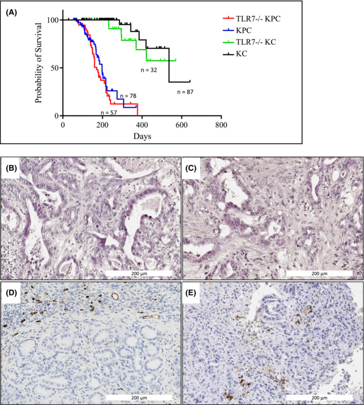
Methods: We used qRT-PCR and immunohistochemistry to asses TLR7 expression in primary patient material and cell lines. Cell viability was analyzed by MTT assay upon incubation with TLR7 agonist/antagonist. Mouse models were used to investigate the role of TLR7 in vivo.

Results: TLR7 is overexpressed in more than 50% of primary human pancreatic ductal adenocarcinoma (PDAC). High TLR7 expression was associated with shorter patient survival, and TLR7 inhibition in cell lines reduced viability in a dose-dependent manner. In contrast, global TLR7 deficiency did not alter survival or overall histopathological tumor features in genetic mouse models of PDAC.

Conclusions: TLR7 may have opposing functions in tumor versus stroma cells. Further work is required to more precisely dissect the roles of TLR7 and its ligands in different populations of epithelial and stromal cells and to understand their relative contributions to tumor progression.


Conflict of interest statement

The authors declare no competing financial interests.

Figures

FIGURE 1
FIGURE 1
Expression of TLR7 in primary human tissues. (A–D) Tissue microarrays (TMAs) of human PDAC (B, D) and corresponding normal (A, C) tissue, were stained against TLR7 (Proteintech #17232‐1‐AP), results showing strong (B) and moderate (D) expression of TLR7 in tumor cells (arrows). Islets of Langerhans (A, asterisk) also stained positive for TLR7. E: Quantification of staining results from TMA sections.
FIGURE 2
FIGURE 2
Correlation of TLR7 expression with patient survival and relative age of cell cultures. (A) Kaplan–Meier curves of overall survival according to TLR7 IHC (censored data are flagged). n = 24; Log‐rank (Mantel‐Cox) test. (B) Correlation of survival and TLR7 expression reveals that all long‐time survivors (<36 months from diagnosis) exhibit weak to moderate TLR7 staining. (C) Q‐RT‐PCR analyses of TLR7 mRNA expression in short‐term cultures upon thawing in comparison to established PDAC cell lines. Expression values are shown relative to the housekeeping gene RPLP0 in each cell line. Bars represent the mean ± SDM of two to five independent experiments.
FIGURE 3
FIGURE 3
TLR7 mediates growth‐stimulatory effects in cancer cells in vitro. (A & B) LON560 (A) and KC623 (B) were treated with different concentrations of IRS‐954/Ctr_ODN [0.5 μM–20 μM] or CL264 [5 μg/mL–0.5 μg/mL or 0.5 μg/mL–0.05 μg/mL]. Cell viability was measured by MTT assay 72 h after treatment. (C) S2‐007, a low‐expressing TLR7 cell line, was treated with 10 μM IRS‐954/Ctr_ODN (concentration with the highest effect in LON560). Cell viability was measured by MTT assay 72 h after treatment. Bars represent the mean ± SDM of at least three independent experiments normalized to control cells (0%‐FCS/ 1%‐FCS). Ctr_ODN, control oligo. (Students t test: *p < 0.05, **p < 0.01, ***p < 0.001).
FIGURE 4
FIGURE 4
Global TLR7 knockout does not attenuate tumor progression in vivo. (A) Kaplan–Meier analyses revealed that TLR7 deficiency did not result in any survival benefit, neither in the KPC nor in the KC mouse model. (B & C) Representative histological sections (H&E staining) of 7‐month‐old WT‐KPC (B) and TLR7−/−‐KPC (C) mice. (D & E) Representative images of CD68 staining for monocytes in 7‐month‐old WT‐KPC (D) and TLR7−/−‐KPC (E) mice.
FIGURE 5
FIGURE 5
Volcano plot of differentially expressed genes. The log2 FC (fold change) indicates the mean expression level for each gene. Positive values denote genes upregulated in TLR7 KO vs. control KPC tumors; negative values denote genes downregulated in TLR7 KO tumors. Each dot represents one gene; red dots denote genes significantly differentially expressed at a false discovery rate (FDR) of 10%.

References

    1. Siegel RL, Miller KD, Jemal A. Cancer statistics, 2020. CA Cancer J Clin. 2020;70(1):7‐30. doi:10.3322/caac.21590 - DOI - PubMed
    1. Singhi AD, Koay EJ, Chari ST, Maitra A. Early detection of pancreatic cancer: opportunities and challenges. Gastroenterology. 2019;156(7):2024‐2040. doi:10.1053/j.gastro.2019.01.259 - DOI - PMC - PubMed
    1. Strobel O, Neoptolemos J, Jager D, Buchler MW. Optimizing the outcomes of pancreatic cancer surgery. Nat Rev Clin Oncol. 2019;16(1):11‐26. doi:10.1038/s41571-018-0112-1 - DOI - PubMed
    1. Gong T, Liu L, Jiang W, Zhou R. DAMP‐sensing receptors in sterile inflammation and inflammatory diseases. Nat Rev Immunol. 2020;20(2):95‐112. doi:10.1038/s41577-019-0215-7 - DOI - PubMed
    1. Jung S, von Thulen T, Yang I, et al. A ribosomal RNA fragment with 2′,3′‐cyclic phosphate and GTP‐binding activity acts as RIG‐I ligand. Nucleic Acids Res. 2020;48(18):10397‐10412. doi:10.1093/nar/gkaa739 - DOI - PMC - PubMed

Publication types

Substances