. 2023 Feb 28;14(1):e0266622.
doi: 10.1128/mbio.02666-22. Epub 2023 Jan 5.

Structural Analyses of a Dominant Cryptosporidium parvum Epitope Presented by H-2Kb Offer New Options To Combat Cryptosporidiosis

Affiliations

Structural Analyses of a Dominant Cryptosporidium parvum Epitope Presented by H-2Kb Offer New Options To Combat Cryptosporidiosis

Yongli Wang et al. mBio. .

Abstract

Cryptosporidium parvum has gained much attention as a major cause of diarrhea in the world, particularly in those with compromised immune systems. The data currently available on how the immune system recognizes C. parvum are growing rapidly, but we lack data on the interactions among host major histocompatibility complex (MHC) diversity and parasitic T-cell epitopes. To identify antigenic epitopes in a murine model, we performed systematic profiling of H-2Kb-restricted peptides by screening the dominant Cryptosporidium antigens. The results revealed that the glycoprotein-derived epitope Gp40/15-SVF9 induced an immunodominant response in C. parvum-recovered C57BL/6 mice, and injection of the cytotoxic-T-lymphocyte (CTL) peptide with the adjuvant activated peptide-specific CD8+ T cells. Notably, the SVF9 epitope was highly conserved across Cryptosporidium hominis, C. parvum, and many other Cryptosporidium species. SVF9 also formed stable peptide-MHC class I (MHC I) complexes with HLA-A*0201, suggesting cross-reactivity between H-2Kb and human MHC I specificities. Crystal structure analyses revealed that the interactions of peptide-MHC surface residues of H-2Kb and HLA-A*0201 are highly conserved. The hydrogen bonds of H-2Kb-SVF9 are similar to those of a dominant epitope presented by HLA-A*0201, which can be recognized by a public human T-cell receptor (TCR). Notably, we found double conformations in position 4 (P4), 5 (P5) of the SVF9 peptide, which showed high flexibility, and multiple peptide conformations generated more molecular surfaces that can potentially be recognized by TCRs. Our findings demonstrate that an immunodominant C. parvum epitope and its homologs from different Cryptosporidium species and subtypes can benefit vaccine development to combat cryptosporidiosis. IMPORTANCE Adaptive immune responses and T lymphocytes have been implicated as important mechanisms of parasite-induced protection. However, the role of CD8+ T lymphocytes in the resolution of C. parvum infection is largely unresolved. Our results revealed that the glycoprotein-derived epitope Gp40/15-SVF9 induced an immunodominant CD8+ T-cell response in C57BL/6 mice. Crystal structure analyses revealed that the interactions of the H-2Kb-SVF9 peptide are similar to those of a dominant epitope presented by HLA-A*0201, which can be recognized by human TCRs. In addition, we found double conformations of the SVF9 peptide, which showed high flexibility and multiple peptide conformations that can potentially be recognized by TCRs.

Keywords: Cryptosporidium parvum; MHC I; T-cell epitope; cell-mediated immunity.


Conflict of interest statement

We declare no conflict of interest.

Figures

FIG 1
FIG 1
Identification of C. parvum-specific CD8+ T-cell epitopes in C57BL/6 mice immunized with oocysts followed by peptide. (A) IFN-γ ELISpot analysis of splenocytes obtained 7 days after C. parvum oocyst and peptide immunization of mice (n = 6). Seven days after the last immunization, cells were restimulated with 8 peptides in vitro. (B) Splenocytes were isolated with a mouse CD8+ T-cell isolation kit (CD8+ T cell) or without depletion (Total splenocytes). The peptide-immunized groups were immunized with 50 μg (100 μL) of either peptide packaged with Freund’s adjuvant in a total volume of 200 μL per mouse. The adjuvant controls received 100 μL of PBS and 100 μL of Freund’s adjuvant, and the naive controls were each injected with 200 μL of PBS. (C) Representative ELISpot wells and splenocytes of CD8+ T cells isolated from mice immunized with oocysts and peptides. Frequencies of IFN-γ+ cells/total CD8+ T cells are indicated. Data represent means ± standard deviations (SD), with significance determined using Student’s t test (**, P < 0.01; ***, P < 0.001).
FIG 2
FIG 2
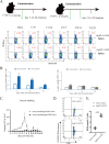
Kinetics of SVF9- and AIF9-specific CD8+ T-cell responses following immunization with C. parvum oocysts. (A) C57BL/6 mice were vaccinated either once or twice with C. parvum (C.p) oocysts, as shown in the schematic diagram. On days 7, 14, and 28 after the last immunization, SVF9- and AIF9-specific CD8+ T-cell responses were quantified in the spleens by peptide stimulation followed by ICS. Representative flow cytometry plots show IFN-γ secretion by CD8+ T cells in the spleens of C. parvum oocyst-immunized mice; 1° represents one immunization with C. parvum oocysts, while 2° represents two immunizations with oocysts. FITC, fluorescein isothiocyanate. (B) Data in panel A presented as bar graphs. Empty bars, 1 immunization; filled bars, 2 immunizations (blue for SVF9 and gray for AIF9). **, P < 0.01 by a Mann-Whitney test for differences between 1 and 2 immunizations. Experiments were performed at least 3 times, with 4 mice per group. (C) Comparative analysis of oocyst shedding intensities in C. parvum oocyst-challenged and rechallenged GKO mice. (D) Tetramer staining for single-cell isolation of SVF9-specific T cells in representative splenocytes stained with the H-2Kb–SVF9 tetramer of CD8+ T cells isolated from oocyst-infected and naive mice. Frequencies of IFN-γ+ cells/total CD8+ T cells are indicated. (E) Data from panel D presented as bar graphs, showing differences in peptide-specific CD8+ T cells between oocyst-infected and naive mouse immunizations. ***, P < 0.001.
FIG 3
FIG 3
Priming with SVF9 peptide elevates protective CTL responses in the small intestine and spleen. (A) Schematic diagram of the methodology. C57BL/6 mice were primed with the SVF9 peptide and challenged with oocysts. Ten days later, the frequencies and quality of SVF9-specific CD8+ T cells in the small intestine and spleen were quantified by ICS. (B) Flow cytometry plots of IFN-γ in spleen cells and IELs. Shown are representative dot plots of CD8+ T-cell IFN-γ production following SVF9 peptide restimulation (10 μg/mL) in vitro. Values indicate the percentages of CD8+ cells within each gate, and data were gated on live CD3+ T cells. (C) Analysis of IFN-γ secretion based on the data in panel B. Bars represent the mean values of the percentages of SVF9-specific CD8+ cells. Individual data are also shown. Differences between oocyst infection and SVF9-vaccinated immunizations are indicated. Data are means ± SD, and significance was determined using one-way analysis of variance (ANOVA) with Dunnett’s multiple-comparison test (***, P < 0.001).
FIG 4
FIG 4
Peptide binding identified by FPLC gel filtration and anion-exchange chromatography with H-2Kb and HLA-A*0201. (A) SVF9 peptide formed stable complexes with H-2Kb and HLA-A*0201 that survived both purifications. Peak 1 corresponds to the aggregated heavy chain, peak 2 corresponds to the correctly refolded complex, and peak 3 corresponds to excess β2m. (Inset) Reduced SDS-PAGE gel (12%) showing peaks 1, 2, and 3. Lane M contains molecular weight markers (in kilodaltons). Both complexes were eluted at NaCl concentrations of 14.0 to 16.0%. The specific peaks are marked with red asterisks. (B) Peptide AIF9 with H-2Kb complexes that tolerated gel filtration but mostly dissociated under anion-exchange chromatography conditions. (C) Nonbinding of the AIF9 peptide with the HLA-A*0201 heavy chain and the negative control from the in vitro refolding assays. The sequences of the peptides are shown above their refolding result graphs. mAU, unit of UV absorbance; mS/cm, unit of anion strength.
FIG 5
FIG 5
Distinct conformations and electronic densities of SVF9 peptides found in H-2Kb M1 and M2. (A and B) Electron density at the 1σ contour level of SVF9 peptides shown in the H-2Kb M1 and M2 structures. The electron density maps are clear and indicate that the two distinct peptide conformations are reliable. (C) Detailed comparison of SVF9 peptides shown as a stick model (green, M1; orange, M2). The mismatch of the two peptides is obvious, especially at P4 Ala and P5 Ile. (D) SVF9 peptide conformations (thick sticks) compared to those of other peptides (thin sticks) with distinct conformations presented by H-2Kb (blue, PBD accession no. 1G7P; pink, accession no. 1FZO; olive, accession no. 1WBZ; cyan, accession no. 2VAB).
FIG 6
FIG 6
Peptide interactions with H-2Kb and HLA-A*0201. (A and B) Hydrogen bonds and water molecules found in H-2Kb–SVF9 M1 (A) and M2 (B). Surface model P1, P2, P3, P6, and P9 residues in the SVF9 peptide are accommodated by the A, B, D, C, and F pockets, respectively; 5 water molecules in M1 form 5 direct bonds. The blue balls represent water molecules. (C) Comparison of peptide conformations and peptide-MHC surface residues of H-2Kb and HLA-A*0201. Detailed MHC surface residues are shown as stick models in M1 (green), M2 (orange), and HLA-A*0201 (cyan). (D) Comparison of the peptide-MHC interactions of P1 Ser, P2 Val, and P3 Phe (the N terminus of PBG) between H-2Kb (green and orange) and HLA-A*0201 (PDB accession no. 7RTR) (blue). (E) Comparison of the interactions of P9 Leu in the F pocket (the C terminus of PBG). The hydrogen bonds between the peptides and the pockets are shown as dashed red (M1), blue (M2), and green (HLA-A*0201) lines.
FIG 7
FIG 7
Compositions of the B, D, and F pockets and interactions of H-2Kb with SVF9. (A) Electrostatic potential of pocket B with the P2 residue (red, negative; blue, positive; gray, neutral), with the SVF9 peptide in green (M1) and orange (M2); (B) hydrogen bonds and van der Waals interactions between peptides and the B pocket of M1 and M2 molecules; (C) electrostatic potential of pocket C with the P6 residue; (D) interactions between P6 and the C pocket; (E) electrostatic potential of pocket F with the P9 residue; (F) interactions between P9 and the F pocket. The residues comprising the pockets and the residues of bound peptides accommodated by the pockets are shown as stick models. The hydrogen bonds between the peptides and the pockets are shown as dashed red (M1) and blue (M2) lines.
FIG 8
FIG 8
Distribution of H-2Kb-restricted nonapeptides in important Cryptosporidium species. Shown are the results of genome-wide scanning for H-2Kb-restricted nonapeptides in C. parvum, C. hominis (C.h), and C. felis. GenBank accession numbers are as follows: ACQ82740.1 (C. hominis), AAL07532.1 (C. parvum), and ABH11920.1 (C. felis) for Gp40/15; XP_666956.1 (C. hominis), AAA28294.1 (C. parvum), and KAF7458128.1 (C. felis) for Cp15; AEJ22864.1 (C. hominis), AAN31184.1 (C. parvum), and KAF7456923.1 (C. felis) for Cp23; AIT12349.1 (C. hominis), AGS42174.1 (C. parvum), and AKM20838.1 (C. felis) for HSP70; CUV04935.1 (C. hominis) and EAK89290.1 (C. parvum) for CSL; and OLQ18067.1 (C. hominis) and AAC98153 (C. parvum) for Gp900. Forty-five nonapeptides derived from the Gp40/15, Cp23, Cp15, HSP70, CSL, and Gp900 proteins were selected based on the modified motif X-(I/V/M/L/A/G/R/S)-X-X-(Y/F/W/L/I/K/N/T)-(F/Y/R/P/N/S/K/H/L/I/V)-X-X-(L/M/V/I/A).
FIG 9
FIG 9
Analyses of contact between human HLA-A*0201 and the public TCR and modeling of the H-2Kb–TCR complex. (A) Contacts between the public TCR and MHC surface residues on the tops of the helices. Models of the structures of the H-2Kb–TCR complex (pink sticks), the HLA-A*0201–TCR complex (blue sticks), H-2Kb surface residues (green sticks), HLA-A*0201 (cyan sticks), and H-2Kb molecule α1 and α2 domains (white cartoon) are shown. The hydrogen bonds and interaction residues are highly conserved, except for Arg155/Gln155 and Arg65/Gln65. (B) Superposition of the HLA-A*0201–YLQ and H-2Kb–SVF9 peptide structures. In comparison with HLA-A*0201 without the TCR (PDB accession no. 7RTD) (pink), the P5(I) residues of the SVF9 peptides (green in M1 and orange in M2) have conformations more similar to those of P5(R) presented by the HLA-A*0201–TCR complex (PDB accession no. 7RTR) (blue). Asp109β and Asp110α in the conserved motif form two hydrogen bonds with the YLQ peptide, also showing the interaction of the CDR3β and CDR3α loops (red) with the peptide. Interactions are also shown in Table S4.

Similar articles

Cited by

References

    1. Feng Y, Ryan UM, Xiao L. 2018. Genetic diversity and population structure of Cryptosporidium. Trends Parasitol 34:997–1011. doi:10.1016/j.pt.2018.07.009. - DOI - PubMed
    1. Checkley W, White AC, Jr, Jaganath D, Arrowood MJ, Chalmers RM, Chen X-M, Fayer R, Griffiths JK, Guerrant RL, Hedstrom L, Huston CD, Kotloff KL, Kang G, Mead JR, Miller M, Petri WA, Jr, Priest JW, Roos DS, Striepen B, Thompson RCA, Ward HD, Van Voorhis WA, Xiao L, Zhu G, Houpt ER. 2015. A review of the global burden, novel diagnostics, therapeutics, and vaccine targets for cryptosporidium. Lancet Infect Dis 15:85–94. doi:10.1016/S1473-3099(14)70772-8. - DOI - PMC - PubMed
    1. Shimelis T, Tassachew Y, Lambiyo T. 2016. Cryptosporidium and other intestinal parasitic infections among HIV patients in southern Ethiopia: significance of improved HIV-related care. Parasit Vectors 9:270. doi:10.1186/s13071-016-1554-x. - DOI - PMC - PubMed
    1. Vinayak S, Pawlowic MC, Sateriale A, Brooks CF, Studstill CJ, Bar-Peled Y, Cipriano MJ, Striepen B. 2015. Genetic modification of the diarrhoeal pathogen Cryptosporidium parvum. Nature 523:477–480. doi:10.1038/nature14651. - DOI - PMC - PubMed
    1. Striepen B. 2013. Parasitic infections: time to tackle cryptosporidiosis. Nature 503:189–191. doi:10.1038/503189a. - DOI - PubMed

Publication types