

Effect of veneering material type and thickness ratio on flexural strength of bi-layered PEEK restorations before and after thermal cycling
- PMID: 36602589
- PMCID: PMC10264476
- DOI: 10.1007/s00784-022-04829-8
Effect of veneering material type and thickness ratio on flexural strength of bi-layered PEEK restorations before and after thermal cycling
Abstract
Objective: The purpose of this study was evaluating the biaxial strength of bi-layered PEEK restorations before and after aging using different veneering materials in different thickness ratios.
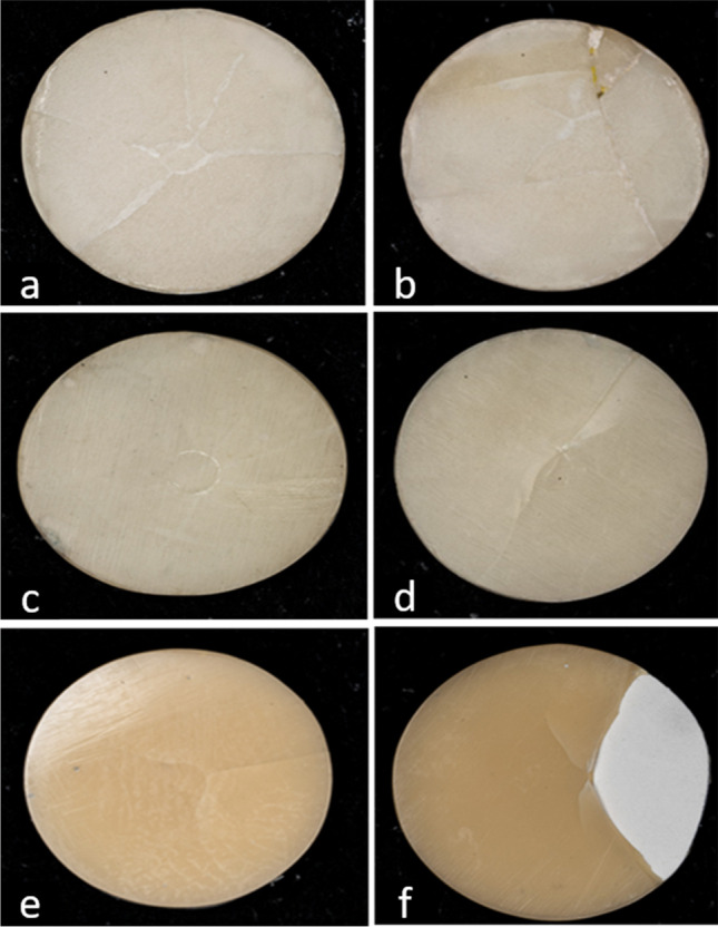
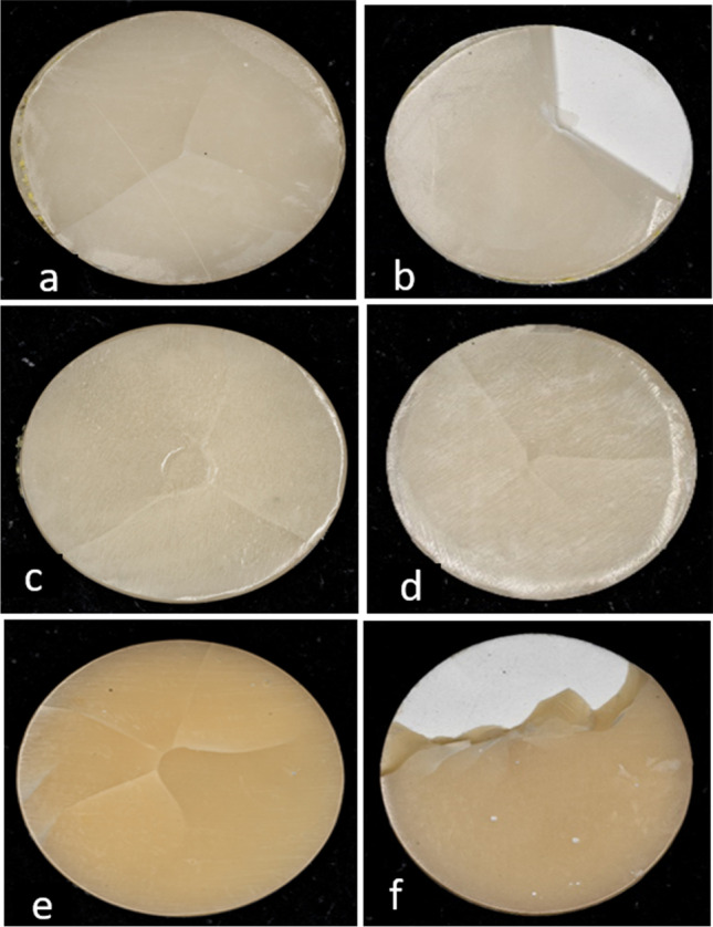
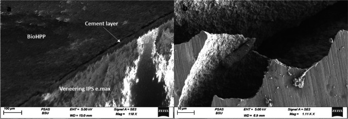
Material and methods: Ninety specimens of thickness 1.5 mm were divided into three groups according to their veneering material. Group (CAD LD): BioHPP discs veneered with CAD milled lithium disilicate (n=30), group (CAD C): BioHPP discs veneered with CAD milled composite (n=30), and group (LC): BioHPP discs veneered with conventionally layered composite (n=30). Each group was subdivided into 3 subgroups (n=10) according to the different thickness ratios between the core and the veneering material (TC:TV). Subgroup 1: TC:TV=1:0.5, subgroup 2: TC:TV=0.7:0.8, and subgroup 3: TC:TV=0.5:1. Half of the specimens of each subgroup were subjected to thermocycling, and the bi-axial flexural strength of all specimens was tested before and after aging. Three-way ANOVA followed by Bonferroni's post hoc test were used for data analysis. The significance level was set at P ≤ 0.05.
Results: Material, thickness ratio, and aging all had a significant effect on biaxial flexural strength. (LC) group had the highest biaxial flexural strength. TC:TV=0.5:1 showed the lowest biaxial flexural strength. All groups showed significant decrease in biaxial flexural strength after aging.
Conclusions: Veneering material for PEEK together with the thickness ratio between the core and veneering material greatly affect the flexural strength of bi-layered restorations. Thermocycling negatively impacts the flexural strength of PEEK bi-layered restorations.
Clinical significance: According to the results of that study, PEEK cores are best veneered with conventionally layered composite with core to veneering thickness ratio being 1:0.5.
Keywords: Flexural strength; PEEK; Thermocycling; Veneering.
© 2023. The Author(s).
Conflict of interest statement
The authors declare no competing interests.
Figures


References
-
- Tartuk B, Anna E, Basaran E. Comparison of the load-bearing capacities of monolithic PEEK, zirconia and hybrid ceramic molar crowns. Meanders Med Dent J. 2019;20:45–50. doi: 10.4274/meandros.galenos.2018.54269. - DOI
-
- Tekin S, Cangül S, Adıgüzel Ö, Deg˘ er Y. Areas for use of PEEK material in dentistry. Int Dent Res. 2018;8(2):84–92. doi: 10.5577/intdentres.2018.vol8.no2.6. - DOI
MeSH terms
Substances
LinkOut - more resources
Full Text Sources
Research Materials
Miscellaneous
