. 2023 Feb 1;141(2):168-175.
doi: 10.1001/jamaophthalmol.2022.5681.

Incidence and Progression of Chorioretinal Folds During Long-Duration Spaceflight

Affiliations

Incidence and Progression of Chorioretinal Folds During Long-Duration Spaceflight

Connor R Ferguson et al. JAMA Ophthalmol. .

Abstract

Importance: The primary contributing factor for development of chorioretinal folds during spaceflight is unknown. Characterizing fold types that develop and tracking their progression may provide insight into the pathophysiology of spaceflight-associated neuro-ocular syndrome and elucidate the risk of fold progression for future exploration-class missions exceeding 12 months in duration.

Objective: To determine the incidence and presentation of chorioretinal folds in long-duration International Space Station crew members and objectively quantify the progression of choroidal folds during spaceflight.

Design, setting, and participants: In this retrospective cohort study, optical coherence tomography scans of the optic nerve head and macula of crew members completing long-duration spaceflight missions were obtained on Earth prior to spaceflight and during flight. A panel of experts examined the scans for the qualitative presence of chorioretinal folds. Peripapillary total retinal thickness was calculated to identify eyes with optic disc edema, and choroidal folds were quantified based on surface roughness within macular and peripapillary regions of interest.

Interventions or exposures: Spaceflight missions ranging 6 to 12 months.

Main outcomes and measures: Incidence of peripapillary wrinkles, retinal folds, and choroidal folds; peripapillary total retinal thickness; and Bruch membrane surface roughness.

Results: A total of 36 crew members were analyzed (mean [SD] age, 46 [6] years; 7 [19%] female). Chorioretinal folds were observed in 12 of 72 eyes (17%; 6 crew members). In eyes with early signs of disc edema, 10 of 42 (24%) had choroidal folds, 4 of 42 (10%) had inner retinal folds, and 2 of 42 (5%) had peripapillary wrinkles. Choroidal folds were observed in all eyes with retinal folds and peripapillary wrinkles. Macular choroidal folds developed in 7 of 12 eyes (4 of 6 crew members) with folds and progressed with mission duration; these folds extended into the fovea in 6 eyes. Circumpapillary choroidal folds developed predominantly superior, nasal, and inferior to the optic nerve head and increased in prevalence and severity with mission duration.

Conclusions and relevance: Choroidal folds were the most common fold type to develop during spaceflight; this differs from reports in idiopathic intracranial hypertension, suggesting differences in the mechanisms underlying fold formation. Quantitative measures demonstrate the development and progression of choroidal folds during weightlessness, and these metrics may help to assess the efficacy of spaceflight-associated neuro-ocular syndrome countermeasures.


Conflict of interest statement

Conflict of Interest Disclosures: Drs Laurie and Macias reported grants from NASA during the conduct of the study. No other disclosures were reported.

Figures

Figure 1.
Figure 1.. Quantification of Bruch Membrane Surface Roughness
Bruch membrane surface layer was used to visualize and quantify the development of choroidal folds and their progression during spaceflight. Bruch membrane layer (red) and internal limiting membrane (blue) were manually segmented on optical coherence tomography images from the vertical block scan centered over the fovea (A) and the radial scan centered over the optic nerve head (B). Inner retinal folds (white arrowhead) and choroidal folds (yellow arrowheads) are marked on each transverse optical coherence tomography image. The subtle retinal folds indicated in panel A are extensions of the prominent inner retinal folds observed in the same crew member in Figure 2B. Bruch membrane surface layer colored height maps were generated to better visualize the pattern and orientation of macular (C) and peripapillary choroidal folds (D) during spaceflight: teal represents no change, while dark blue and yellow represent posterior and anterior displacement, respectively. The change in Bruch membrane macular surface roughness was quantified within 3 adjacent 1 × 5-mm regions to track the successive progression of choroidal folds spanning from the peripapillary region (C: fovea–2 mm ROI) toward the fovea region (C: fovea ROI) during spaceflight. Peripapillary surface roughness was calculated within the nasal, superior, temporal, and inferior quadrants of an elliptical region of interest within 500 to 1000 μm of Bruch membrane opening (D).
Figure 2.
Figure 2.. Distribution of Choroidal Folds, Retinal Folds, and Peripapillary Wrinkles in International Space Station (ISS) Crew Members
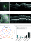
A, Example optical coherence tomography (OCT) image indicating a region of peripapillary wrinkles (white arrowheads). B, Example OCT image indicating region of inner retinal folds (white arrowhead) and choroidal folds (yellow arrowhead). In panels A and B, the infrared image includes the OCT scan pattern location (green lines) with the bold line representing the OCT image to the right. C, Of the 36 participants in this study, all 6 individuals who presented with folds during spaceflight demonstrated bilateral choroidal folds. Within these 6 individuals, 8 eyes had only choroidal folds, 2 eyes had both choroidal folds and inner retinal folds, and 2 eyes had choroidal folds, inner retinal folds, and peripapillary wrinkles. D, The prevalence of each fold type within ISS crew member eyes demonstrating the earliest signs of optic disc edema (data presented here) differed from the prevalence of each fold type within eyes of patients with idiopathic intracranial hypertension (IIH) demonstrating papilledema., Within the 42 eyes that showed signs of developing optic disc edema during spaceflight, 2 eyes (5%) had peripapillary wrinkles, 4 (10%) had inner retinal folds, and 10 (24%) had choroidal folds. As reported by Sibony et al,, of 125 study eyes with papilledema, 58 (46%) had peripapillary wrinkles, 59 (47%) had inner retinal folds, 25 (20%) had outer retinal folds, and 13 (10%) had choroidal folds. TRT indicates total retinal thickness.
Figure 3.
Figure 3.. Progression of Macular Choroidal Folds in International Space Station Crew Members During Spaceflight
Compared with preflight, macular Bruch membrane surface roughness increases with spaceflight duration in crew members with macular choroidal folds. Seven study eyes from 4 individual crew members demonstrated choroidal folds in the 3 macular regions of interest (see Figure 1C for locations) during spaceflight. Each individual crew member is represented by a different symbol, which are consistent across figures. Orange symbols represent data from the right eye and blue symbols represent data from the left eye. A meaningful increase in surface roughness was determined when the change compared with preflight exceeded a 2.3-μm threshold (shaded region).
Figure 4.
Figure 4.. Progression of Peripapillary Choroidal Folds in International Space Station Crew Members During Spaceflight
Peripapillary Bruch membrane surface roughness increased nasal, superior, and inferior to the optic nerve head but not temporally. Mean Bruch membrane surface roughness was quantified in an annular region circumscribing the optic nerve head within 500 μm to 1000 μm of Bruch membrane opening. Each individual crew member is represented by a different symbol which are consistent across figures. Orange symbols represent data from the right eye and blue symbols represent data from the left eye. A meaningful increase in surface roughness was determined when the change compared with preflight exceeded a 2.8-μm threshold (shaded region).

Comment in

References

    1. Mader TH, Gibson CR, Pass AF, et al. . Optic disc edema, globe flattening, choroidal folds, and hyperopic shifts observed in astronauts after long-duration space flight. Ophthalmology. 2011;118(10):2058-2069. doi:10.1016/j.ophtha.2011.06.021 - DOI - PubMed
    1. Mader TH, Gibson CR, Barratt MR, et al. . Persistent globe flattening in astronauts following long-duration spaceflight. Neuroophthalmology. 2020;45(1):29-35. doi:10.1080/01658107.2020.1791189 - DOI - PMC - PubMed
    1. Mader TH, Gibson CR, Pass AF, et al. . Optic disc edema in an astronaut after repeat long-duration space flight. J Neuroophthalmol. 2013;33(3):249-255. doi:10.1097/WNO.0b013e31829b41a6 - DOI - PubMed
    1. Mader TH, Gibson CR, Otto CA, et al. . Persistent asymmetric optic disc swelling after long-duration space flight: implications for pathogenesis. J Neuroophthalmol. 2017;37(2):133-139. doi:10.1097/WNO.0000000000000467 - DOI - PubMed
    1. Patel N, Pass A, Mason S, Gibson CR, Otto C. Optical coherence tomography analysis of the optic nerve head and surrounding structures in long-duration International Space Station astronauts. JAMA Ophthalmol. 2018;136(2):193-200. doi:10.1001/jamaophthalmol.2017.6226 - DOI - PubMed

Publication types

Supplementary concepts